

Page snapshot: Introduction to the structure of the grass plant, including the morphology (form) and anatomy (internal structure) of stems, roots, and leaves. Also covers the organization of the reproductive structures.
Topics covered on this page: Introduction; Stems; Stem morphology; Stem anatomy; Roots; Root morphology; Root anatomy; Leaves; Leaf morphology; Leaf anatomy; Reproductive structures; Inflorescences, spikelets, and florets; Fruits; Resources.
Credits: Funded by the National Science Foundation. Any opinions, findings, and conclusions or recommendations expressed in this material are those of the author(s) and do not necessarily reflect the views of the National Science Foundation.This page incorporates content prepared for a revised manuscript of the Teacher-Friendly Guide to the Evolution Maize (Carlyn Buckler, Dhyan Palanichamy, and Andrielle Swaby, 2019). Additional content and revisions by Elizabeth J. Hermsen (2022).
Updates: Page last updated January 11, 2023.
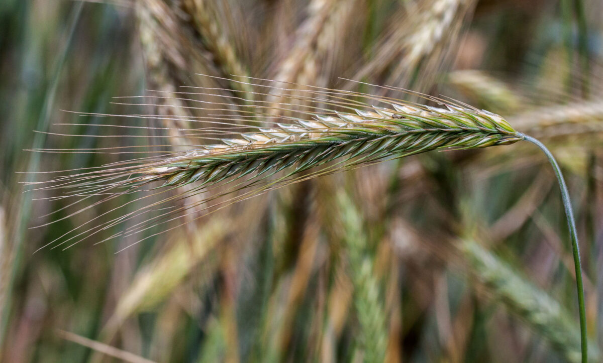
Image above: Inflorescence of rye (Secale cereale). Photo by Fischer.H (Wikimedia Commons, Creative Commons Attribution-ShareAlike 4.0 International license, image cropped and resized).
Introduction
The purpose of this page is to introduce you to the basic structure of the grass plant. Generally speaking, grass plants are organized like other flowering plants. They have three types of vegetative (non-reproductive) organs. These are the stem, which supports the leaves and in grasses may also help the plant spread horizontally; the root, the anchoring and absorptive organ that takes up water and some nutrients; and the leaf, the major photosynthetic organ. Branching in grasses occurs by means of buds that form on the sides of the stems, typically in the site of a leaf axil (the angle between the stem and the upper, or adaxial, side of the leaf). Grasses are typical of monocots in having parallel leaf venation and fibrous root systems. Like other monocots, they cannot produce true wood.
The reproductive unit of a grass is a floret (a tiny flower), and florets are typically arranged in groups called spikelets, which are themselves grouped into larger structures called inflorescences. Grass flowers are very different than what we may think of when we picture a flower. They have no petals and are not very eye-catching. Nevertheless, they produce pollen and ovules (immature seeds), structures that carry out sexual reproduction in the plant.
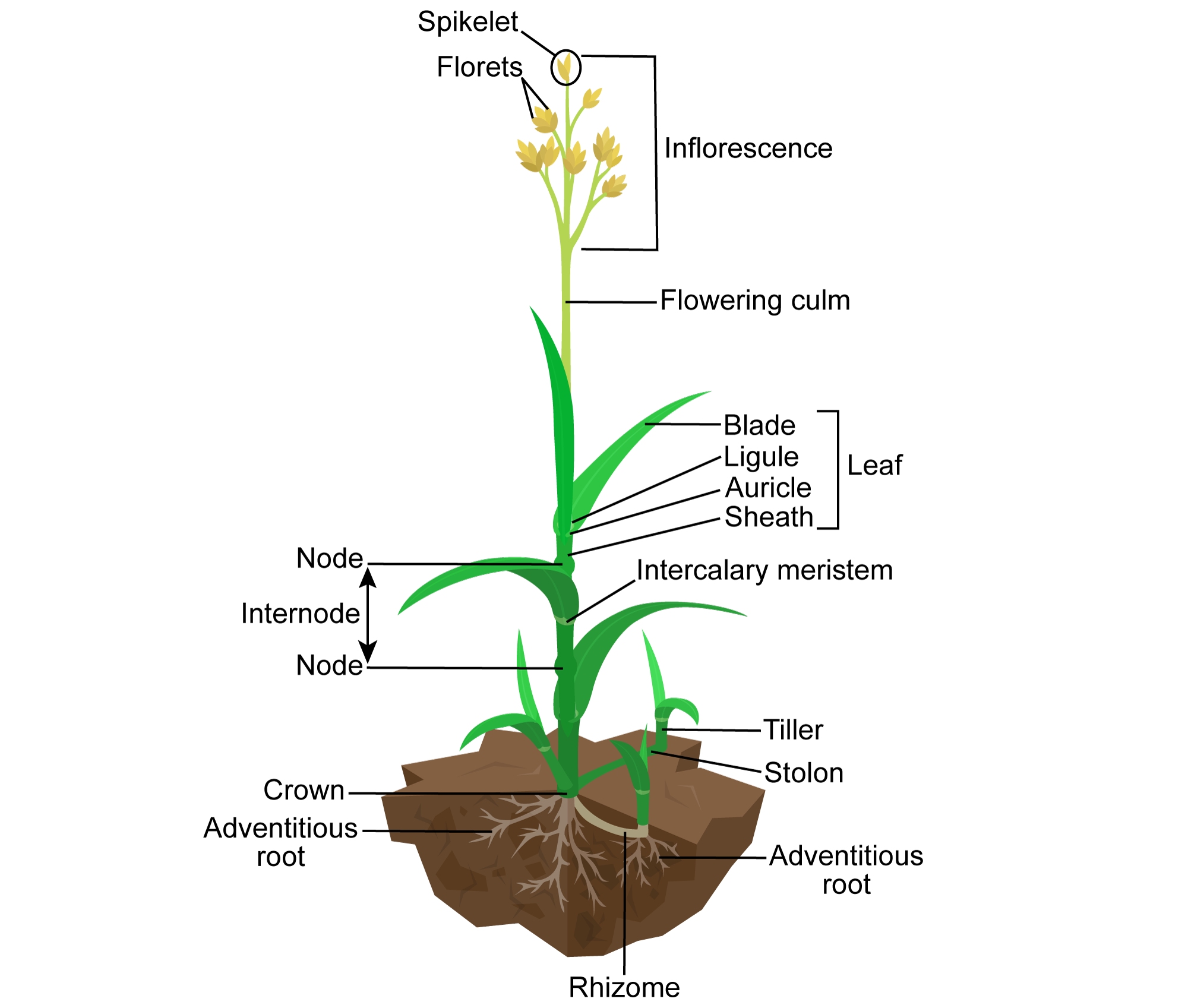
Labeled diagram showing the parts of a grass. Diagram modified from a diagram by Kelvinsong (Wikimedia Commons, Creative Commons Attribution 3.0 Unported license, image modified from original).
Stems
Stem morphology (form)
The stems of grasses, called culms or stalks, are generally cylindrical (circular in cross section). They do not commonly branch above the ground. Daughter shoots, called tillers, may sprout from the base of the stalk. They may also sprout from stolons, stems that creep along the ground for a short distance, or from rhizomes, stems that grow horizontally below the ground. The base of the plant at ground level is called the crown. Most grasses can send up new tillers from the crown if the original stem is cut.
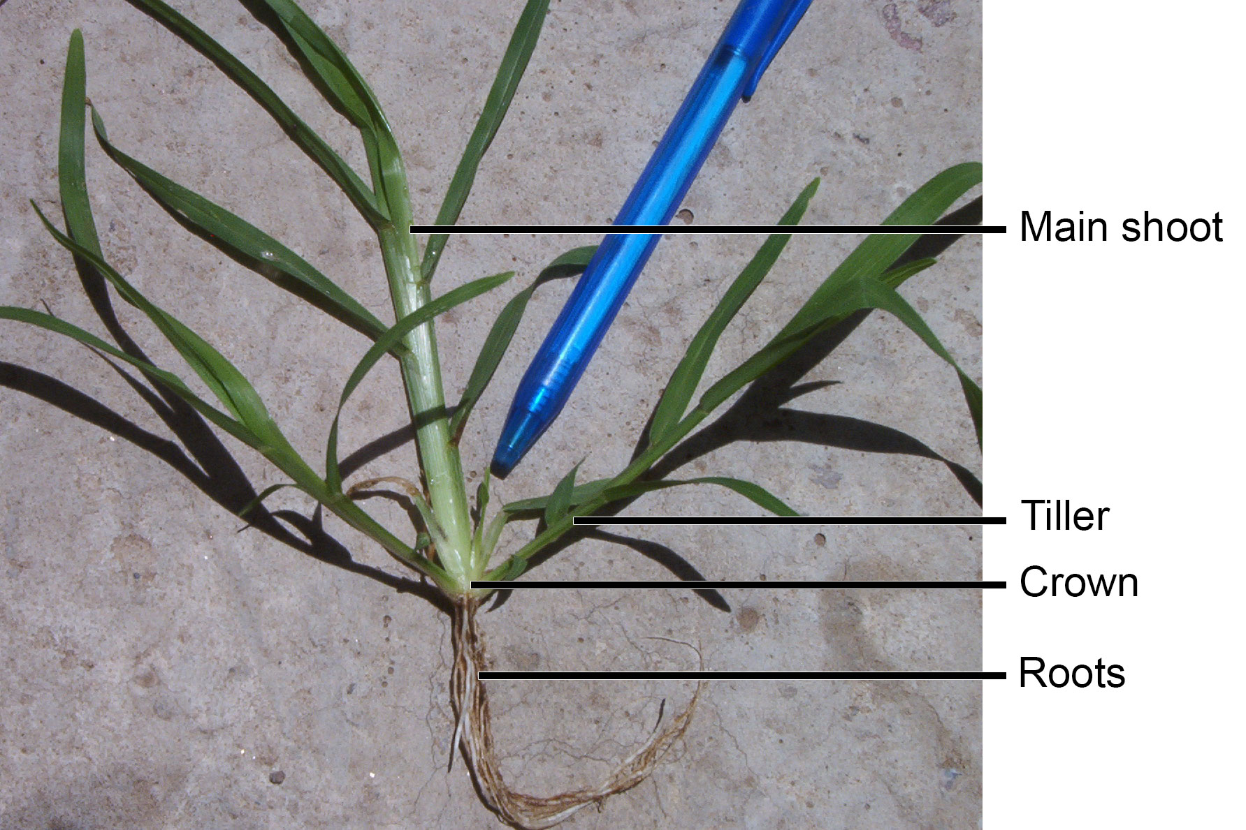
Base of a grass plant showing a main stem flanked by tillers. Photo by RoRo (Wikimedia Commons, Creative Commons CC0 1.0 Universal/public domain dedication).
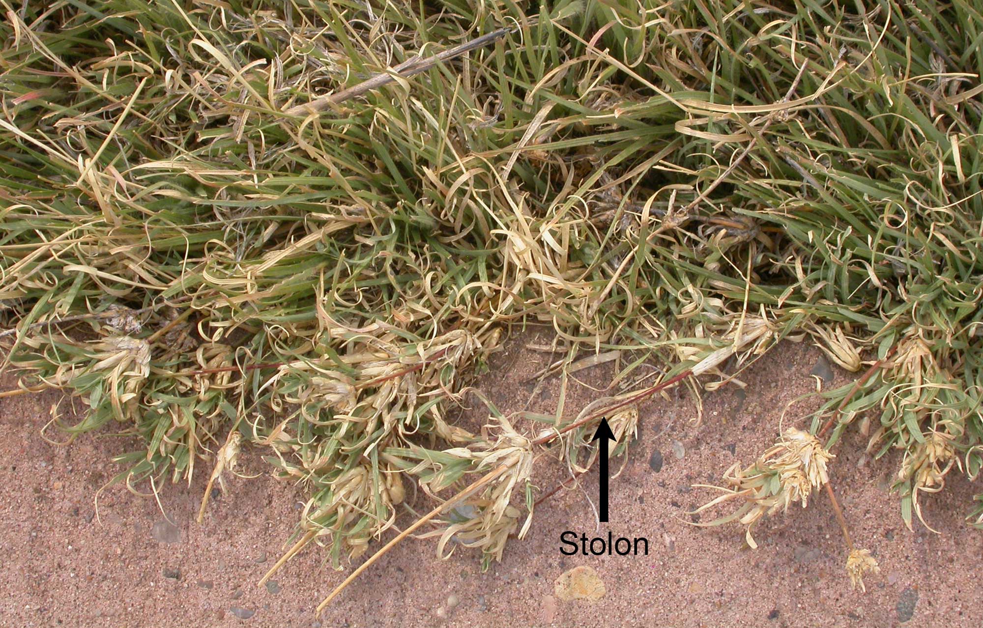
Buffalo grass (Buchloe dactyloides) with stolons. Photo by Matt Lavin (flickr, Creative Commons Attribution-ShareAlike 2.0 Generic license, image cropped, resized, labeled).
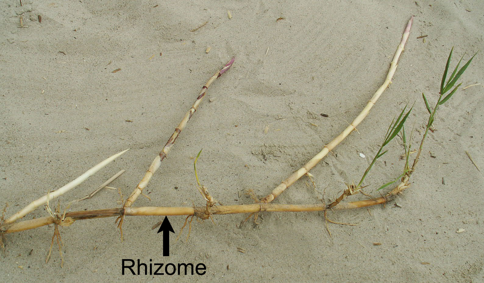
Common reed (Phragmites australis) with branching rhizome. Photo by Kenraiz (Wikimedia Commons, Creative Commons Attribution-ShareAlike 4.0 International license, image labeled).
The stem of a grass is punctuated by nodes. A node is a place where a leaf attaches to a stem. In grasses, nodes are often swollen; because of their swollen nodes, grass stems are sometimes described as having jointed stems. Intervals of stem between nodes are known as internodes.
Note: Be careful in identifying grass nodes! Grasses have sheathing leaves, meaning that the base of the leaf forms a sheath that clasps the stem, whereas the leaf blade is free. The node is found at the base of the sheath. For more discussion, see the section on leaves below.
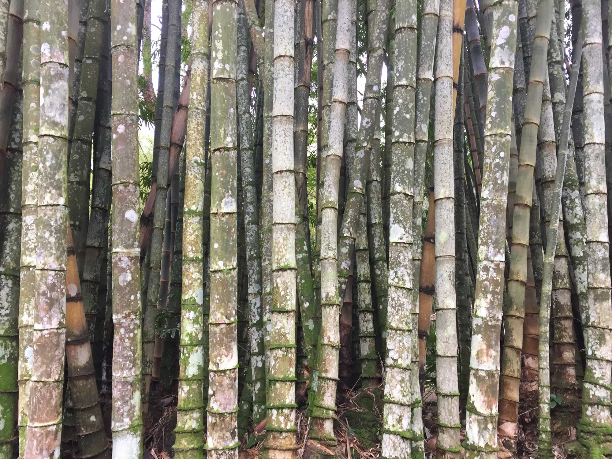
It is easy to find notes on bamboo stems, because the nodes look like grooves around the stem. Nodes are still considered nodes even if the leaves fall off the plant. Photo by PLBechly (Wikimedia Commons, Creative Commons Attribution-ShareAlike 4.0 International license, image resized).
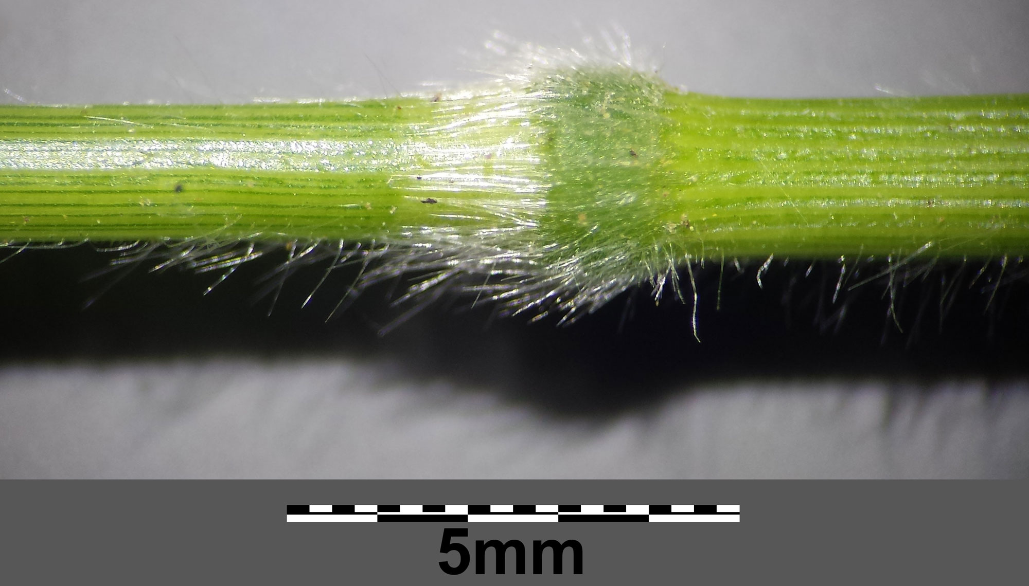
Node (swollen area with longer, denser hairs) of false brome (Brachypodium sylvaticum subspecies sylvaticum). In this photo, the apex of the plant would be toward the left, the base toward the right. Photo by Stefan.Iefnaer (Wikimedia Commons, Creative Commons Attribution-ShareAlike 4.0 International license, images cropped, labeled, and resized).
Stem anatomy (internal structure)
Grass stems are frequently hollow in the internodes and solid at the nodes; however some grasses, like maize (Zea), have a solid core in both the nodal and internodal regions. The outside of the stem is covered by a layer of cells known as the epidermis. The inside of the stem consists of vascular bundles (strands of vascular tissue, meaning water-conducting xylem and food-conducting phloem) that are surrounded by ground tissue (tissue within the epidermis and surrounding the vascular tissue) that is often made up of thin-walled cells known as parenchyma cells. The vascular bundles are either scattered randomly or arranged in two rings within the ground tissue. Each vascular bundle typically has a bundle cap made up of thick-walled, elongated cells called fibers.
The vascular bundles of grasses cannot transition to secondary growth, or the type of growth that produces wood. Thus, grasses are always non-woody plants. (Yes, even bamboo!)
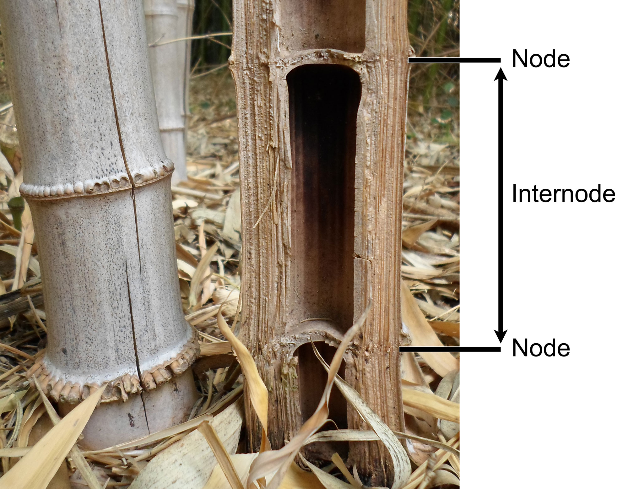
Bamboo stem cut longitudinally showing hollow internodes and solid diaphragms of tissue at the nodes. Photo by Alain Van den Hende (Wikimedia Commons, Creative Commons Attribution-ShareAlike 4.0 International license, image modified from original).
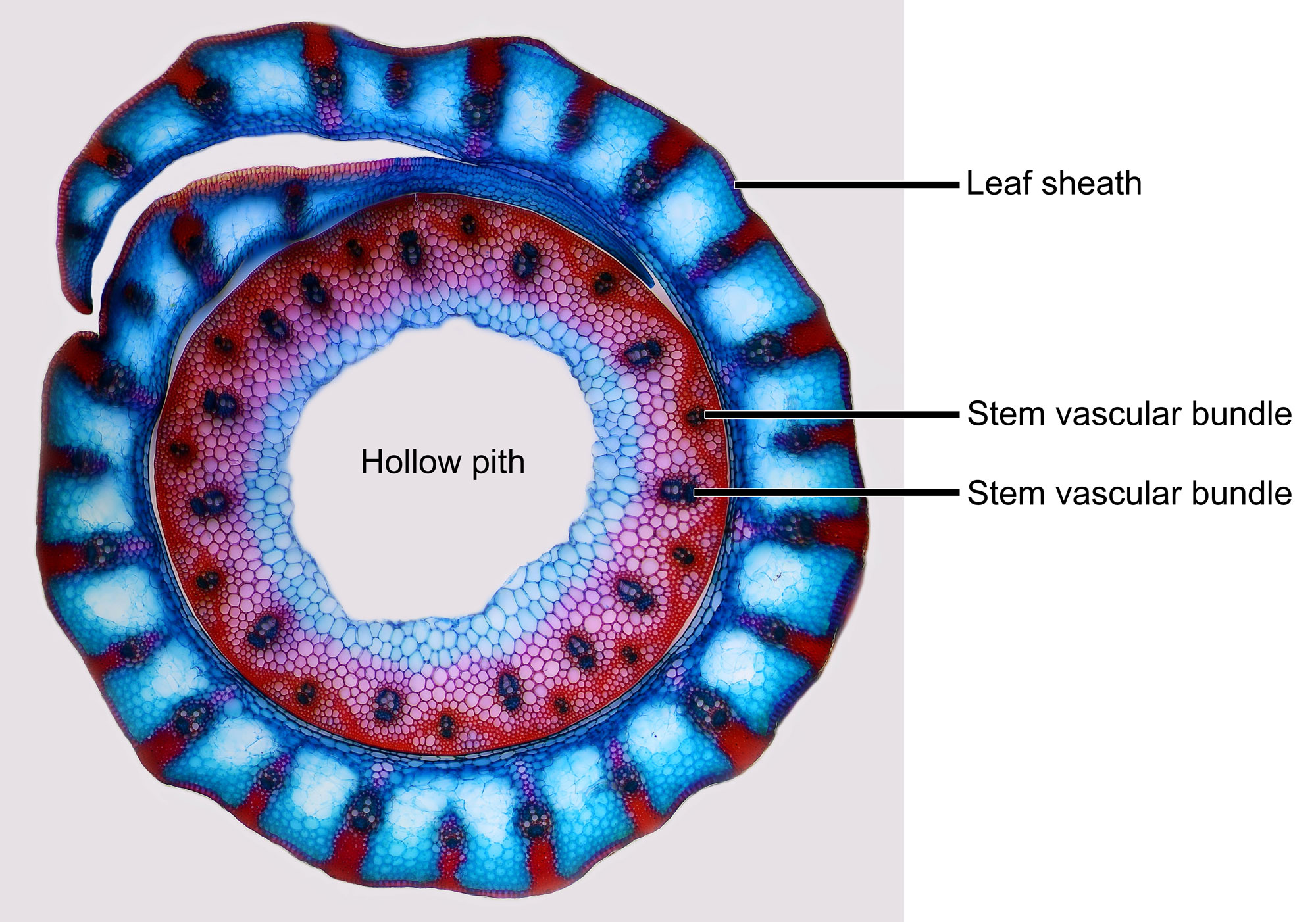
Cross-section of a rye (Secale cereale) stem surrounded by a leaf sheath in an internodal region. The stem has a hollow center and two rings of vascular bundles (water- and food-conducting tissue) near the periphery. A leaf sheath surrounds the stem. Photo by Anatoly Mikhaltsov (Wikimedia Commons, Creative Commons Attribution 4.0 International, image modified from original).

Cross-section of a maize (Zea mays) stem. The vascular bundles (water- and food-conducting tissue) are scattered throughout ground tissue in the stem. Photo by John Houseman and Matthew Ford (Wikimedia Commons, Creative Commons Attribution-ShareAlike 4.0 International license, image modified from original).
Roots
Root morphology
Grasses have a fibrous root system. This root system does not develop from the radicle, or the root of the grass embryo. Instead, it is made up of adventitious roots, or roots that grow from the stem. The fibrous root systems of grasses differ in size and depth depending on environmental and genetic factors as well as the amount of grazing or mowing that occurs during the plant’s lifetime. Unlike stems, roots are not organized into nodes and internodes.
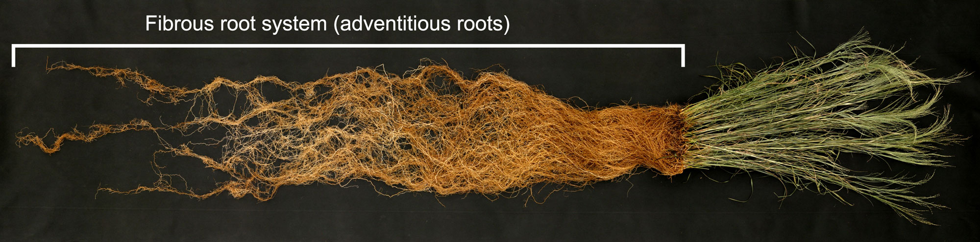
Switchgrass (Panicum virgatum) showing fibrous root system made up of similarly sized adventitious roots. Photo by Lee R. Dehaan (Wikimedia Commons, Creative Commons Attribution 3.0 Unported license, image cropped, resized, label added).
Root anatomy
The grass root is covered by an outer epidermis. Beneath the epidermis is a region of ground tissue known as the cortex. Ground tissue may also occur in the center of the root, forming a pith. The vascular tissues may be organized in a ring between the cortex and pith, although the vascular anatomy of grass roots varies.
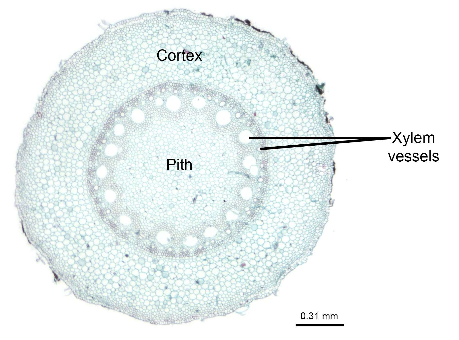
Cross-section of a maize (Zea mays) root. The vascular bundles (water- and food-conducting tissue) are arranged in a ring. The ring of vascular tissue is most easily identified by locating the large xylem vessels (water-conducting cells). Photo by John Houseman and Matthew Ford (Wikimedia Commons, Creative Commons Attribution-ShareAlike 4.0 International license, image modified from original).
Leaves
Leaf morphology
Grass leaves are alternate and distichous in arrangement. This means that one leaf is attached at each node, and the leaves form two vertical rows on the stem. There are two main parts of a grass leaf: the blade and the sheath. The sheath, extending from the node toward the tip of the stem, surrounds the stem like a tube. Farther up the leaf is the blade, which is the part of the leaf that does not surround the stem but instead extends outward to intercept sunlight. The blade usually tapers to a pointed or blunt tip.
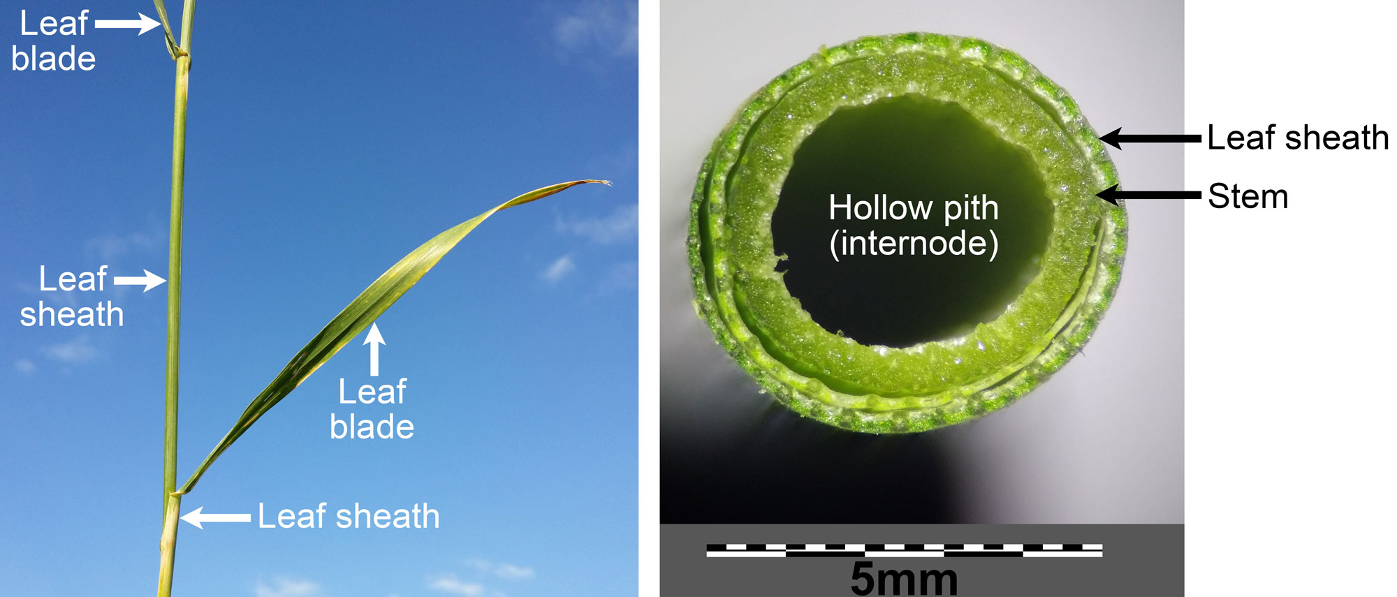
Leaves of spelt (Triticum spelta), a type of wheat. Left: Photo of culm (shoot) of a spelt plant showing leaves with blades and sheaths. Right: Cross section of a spelt culm (shoot) showing the stem with a hollow pit in an internodal region and the encircling leaf sheath. Left photo and right photo by Stefan.Iefnaer (Wikimedia Commons, Creative Commons Attribution-ShareAlike 4.0 International license, images cropped, labeled, and resized).
The ligule is a structure at the junction of the sheath and the blade. It is a membranous flap of tissue or a ring of tiny hairs on the upper side (adaxial side) of the leaf. The ligule clasps the stem, preventing dirt and water from entering the sheath. The outside (also known as the abaxial side, or the side facing away from the stem) of the leaf at the junction between the sheath and blade is called the collar. The collar is often distinctive and can be used to identify different grass species. Some grasses also have auricles, small projections at the base of the blade that may help to prevent the blade from being torn away from the stem.
Grass leaves have long, parallel veins. Veins are strands of vascular tissue. The large parallel veins are connected by minute transverse veins. The parallel veins converge where the leaf blade tapers at its tip. The midvein is the central vein of the leaf.
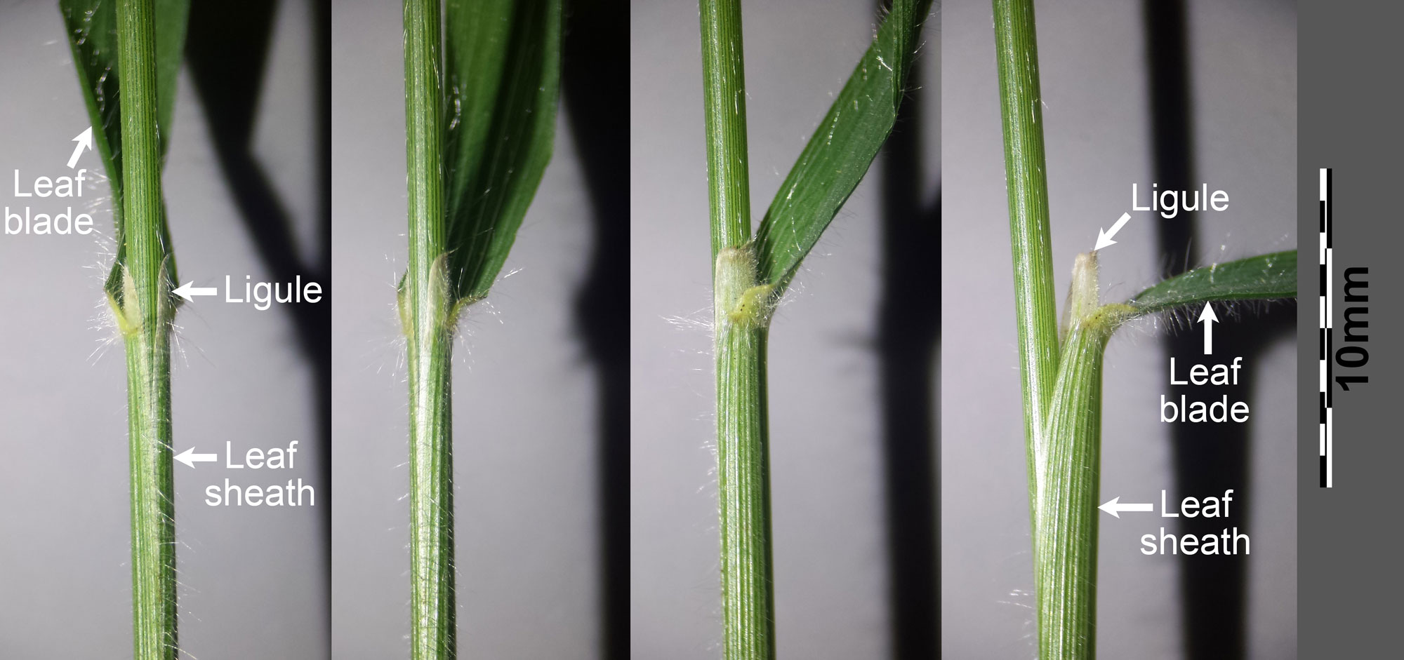
Four views of the leaf blade, leaf sheath, and ligule of false brome (Brachypodium sylvaticum subspecies sylvaticum). Photo by Stefan.Iefnaer (Wikimedia Commons, Creative Commons Attribution-ShareAlike 4.0 International license, images cropped, labeled, and resized).
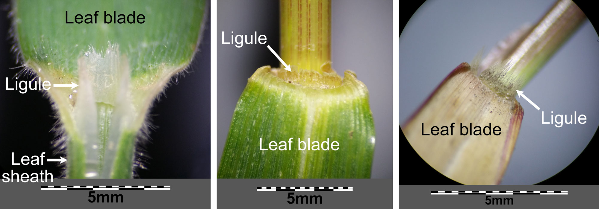
Ligules on the leaves of difference grasses. Left: Velvet grass (Holcus lanatus). Center: Spelt (Triticum spelta). Right: Fall panicgrass (Panicum dichotomiflorum). All photos by Stefan.Iefnaer (Wikimedia Commons, Creative Commons Attribution-ShareAlike 4.0 International license, images cropped, labeled, and resized).
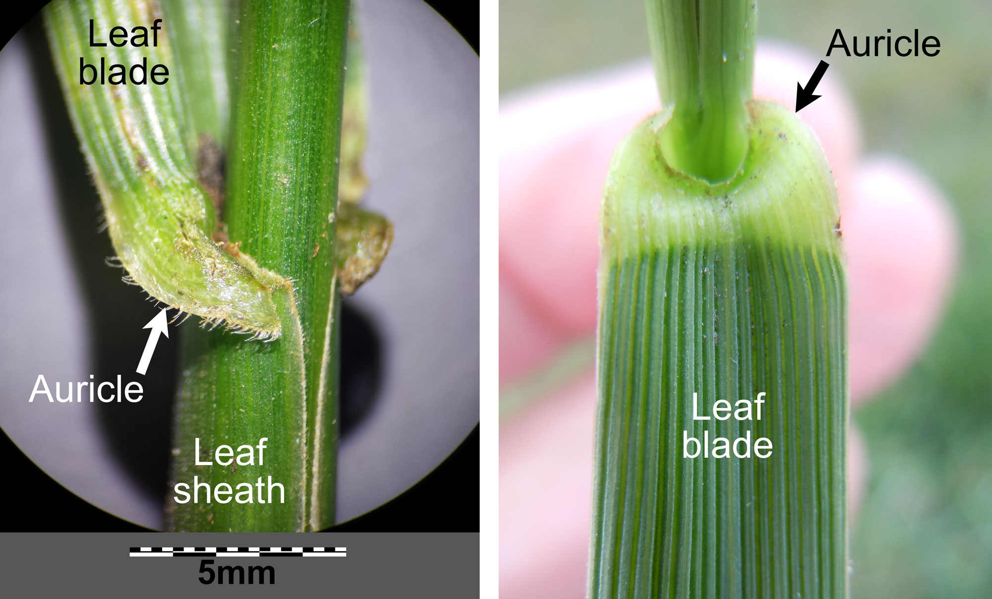
Auricle at the base of the leaf blade in tall fescue (Festuca arundinaceus). Left: Leaf viewed from the side. Right: Leaf viewed from above (adaxial or upper surface of the leaf blade shown). Left photo by Stefan.Iefnaer (Wikimedia Commons, Creative Commons Attribution-ShareAlike 4.0 International license) and right photo by Tim1357 (Wikimedia Commons, Creative Commons Attribution-ShareAlike 2.0 Generic license), images cropped, labeled, and resized).
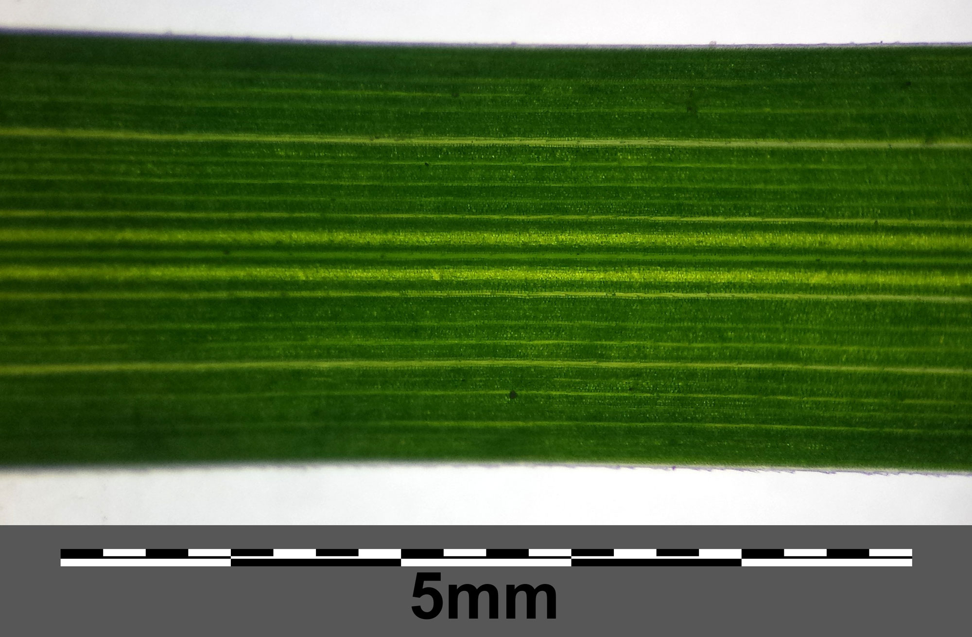
Detail of wood bluegrass (Pea nemoralis) leaf with parallel venation. Photo by Stefan.Iefnaer (Wikimedia Commons, Creative Commons Attribution-ShareAlike 4.0 International license, image cropped and resized).
Leaf anatomy
A grass leaf in cross section is typically thin, with an upper and lower epidermis surrounding a series of roughly circular vascular bundles (veins) embedded in ground tissue called mesophyll. The cells of the mesophyll contain chloroplasts (membrane-bound structures containing green pigment) that carry out photosynthesis.
Leaf epidermis
The grass leaf epidermis is made up of rows of cells that parallel the length (the long axis) of the leaf. These include elongated long cells, short cells that are about the same size in length and width, large bulliform cells, and the cells of the stomatal apparatuses that surround the stomata (pores) in the epidermis.
Much of the epidermis is made up of long cells. The walls on the long sides of the long cells may be sinuous, giving them a puzzle-piece like appearance. Short cells occur singly or in short rows, with the long cells between them. The short cells include silica cells that contain silica bodies and cork cells that have suberin (a fatty compound) in their walls. In some grasses, large bulliform cells may occur in rows in the upper epidermis. These specialized cells can cause the leaf to fold or curl up in hot, dry conditions, helping to prevent water loss.
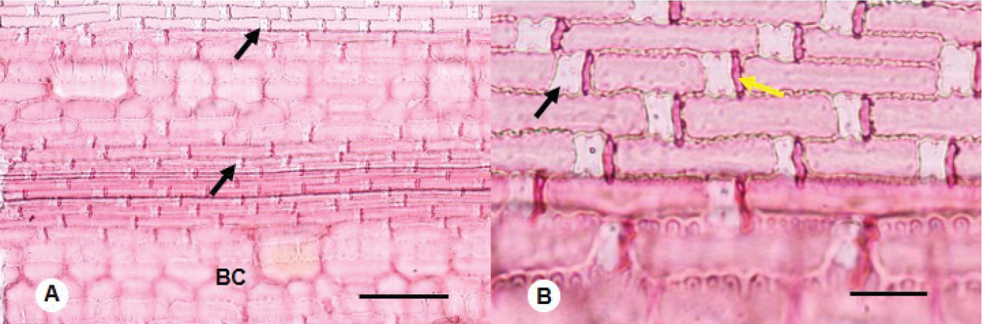
Epidermis of a herbaceous bamboo (Buergersiochloa bambusoides) at two magnifications under a microscope, treated with a stain. Left (A): Black arrows point to examples of silica cells (short cells) alternating with long cells. BC indicates bulliform cells. Scale bar = 100 microns (0.1 millimeters). Right (B): Long cells with short cells between them. The black arrow indicates an example of a silica cell, the yellow arrow indicates an example of a cork cell. The large, out-of-focus cells at the bottom of the image are bulliform cells. Scale bar = 25 microns (0.025 millimeters). Source: From figure 1 in Lima et al. (2021) Phytokeys 172: 135-143 (Creative Commons Attribution 4.0 International license, image cropped).
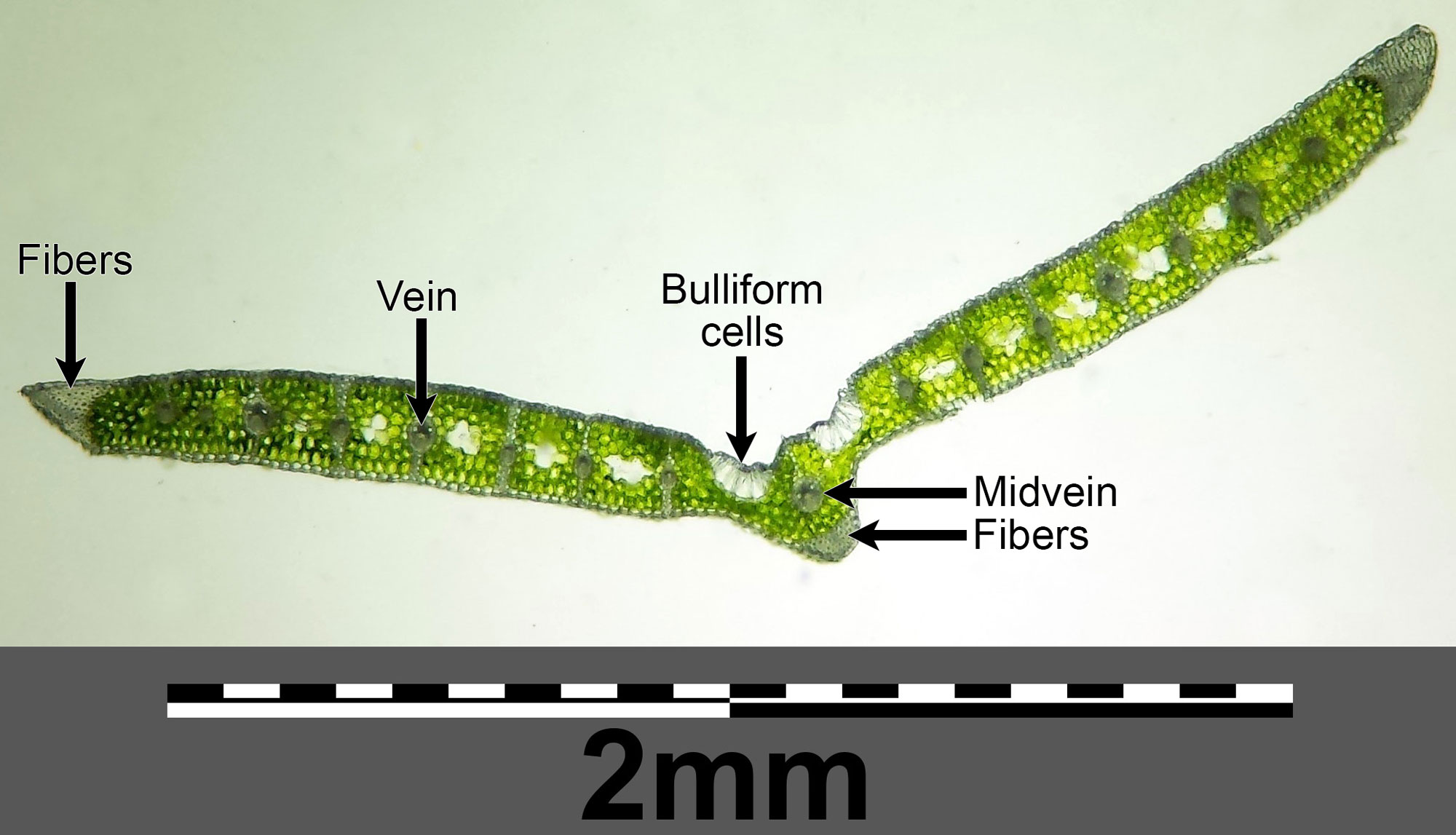
Cross-section of a leaf of blue moor grass (Sesleria caerulea) showing two groups of bulliform cells near the midvein. Groups of fibers help provide strength to the leaf. A fiber is a type of elongated, thick-walled cell that is typically dead when fully mature. Photo by Stefan.Iefnaer (Wikimedia Commons, Creative Commons Attribution-ShareAlike 4.0 International license, image labeled, cropped, and resized).
The epidermis is punctured by stomata that exchange gases. Each stoma is surrounded by a stomatal apparatus made up of guard cells and subsidiary cells. The guard cells open and close the stoma. While in many plants the guard cells of the stomata are kidney-shaped, in grasses they are dumbbell-shaped. Each guard cell is flanked by a subsidiary cell. Subsidiary cells in grasses may be triangular, dome-shaped, or parallel to the guard cells. Long cells with concave end walls occur at either end of each stomatal apparatus; these are sometimes called interstomatal cells.
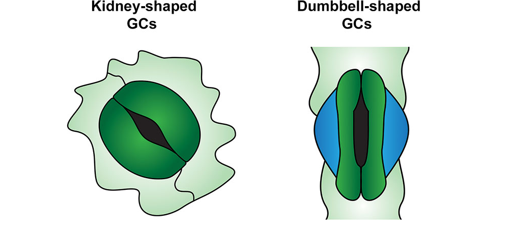
Drawing showing typical, kidney-shaped guard cells (left) and dumbbell-shaped guard cells of a grass (right). The blue cells on either side of the grass guard cells are subsidiary cells. Source: From Figure 1 in Buckley et al. (2020) Frontiers in Plant Science (Creative Commons Attribution 4.0 International license, image cropped).
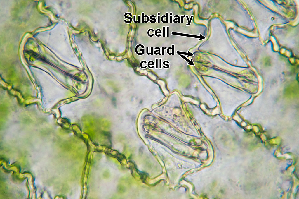
Microscopic image of maize (corn, Zea mays) epidermis showing three stomata with guard cells and subsidiary cells. In this image, the stomata are closed. The cells between the stomata are interstomatal cells, a type of long cell (although in this image, relatively short). Photo by Umberto Salvagnin (flickr, Creative Commons Attribution 2.0 Generic license, image labeled).
Leaf vascular bundles (veins)
In cross section, the vascular bundles in a leaf are arranged more or less in a line. Xylem (water-conducting tissue) occurs toward the upper side of each vascular bundle, and phloem (food-conducting tissue) toward the lower side. Thick-walled cells may extend from the vascular bundles toward the upper and lower sides of the leaf to provide support.
The vascular tissue of each vascular bundle is surrounded by one or two rings of cells that make up a bundle sheath. Most grasses have a two-layered bundle sheath; the outer ring of cells is called the parenchymatous sheath and the inner ring of cells is the mestome sheath. The cells of the mesotome sheath may have thick walls impregnated with a fatty material called suberin, which is waterproof. Some C4 grasses have a single-layered bundle sheath that lacks a mestome sheath.
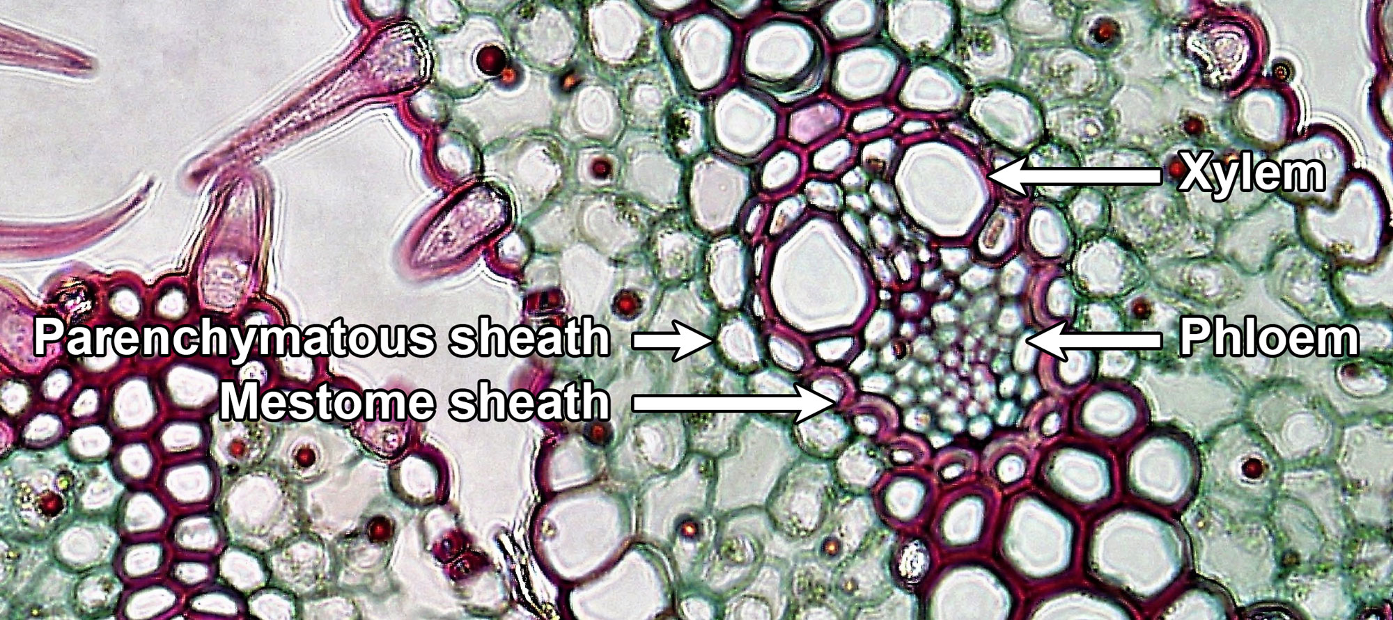
Fixed and stained cross section of a leaf of beachgrass (Ammophila) showing a vascular bundle encircled by a double-layered bundle sheath. The thick, pink-stained inner walls on the cells of the mesotome sheath contain suberin. Beachgrass is a C3 grass. Photo by Berkshire Community College (flickr, CC0 1.0 Univerisal/public domain dedication).
In C4 grasses, the cells of the parenchymatous sheath or bundle sheath (if only a single layer of cells is present) have chloroplasts. The chloroplasts are needed in the bundle sheath cells of C4 grasses because carbon is fixed (incorporated into sugars) in these cells, a process that takes place in the chloroplasts. The bundle sheath cells of C3 grasses have few chloroplasts, because the entire process of photosynthesis can take place in the mesophyll cells in these plants.
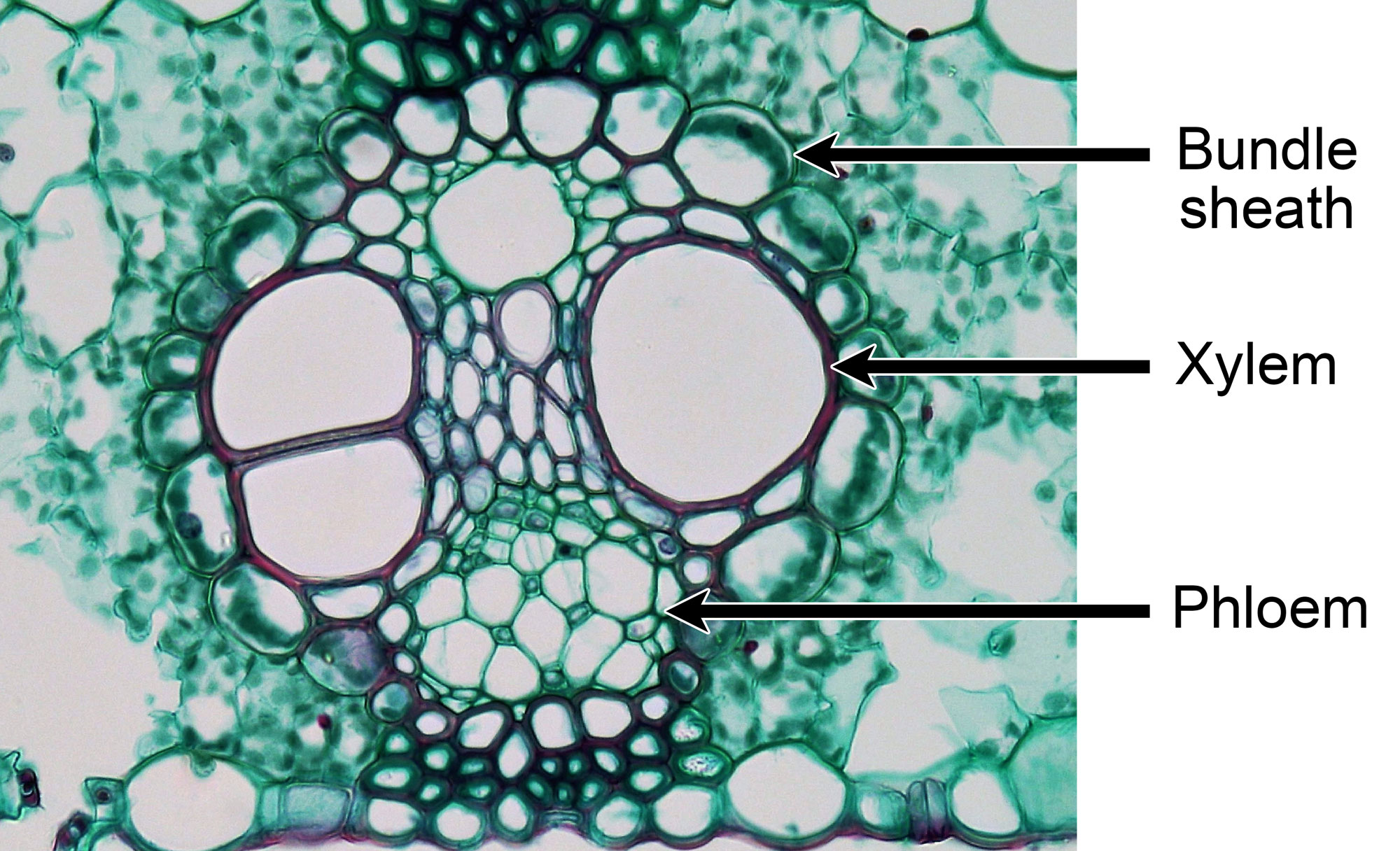
Fixed and stained cross section of a leaf of maize (corn, Zea mays) showing a large vascular bundle encircled by a single-layered bundle sheath with cells containing chloroplasts. Maize is a C4 grass. Photo by Berkshire Community College (flickr, CC0 1.0 Univerisal/public domain dedication).
Mesophyll
The arrangement of cells in the mesophyll, a tissue made up of thin-walled parenchyma cells, varies depending on the type of grass. In C3 grasses, the mesophyll that looks similar throughout the leaf. In C4 grasses, the mesophyll cells are arranged in rings around the vascular bundles.
The anatomical differences between C3 and C4 grasses have to do with differences in the process of photosynthesis between these two types of plants. In C3 plants, photosynthesis takes place in the mesophyll cells. In C4 plants, photosynthetic reactions begin in the mesophyll cells and end in the bundle sheath cells.
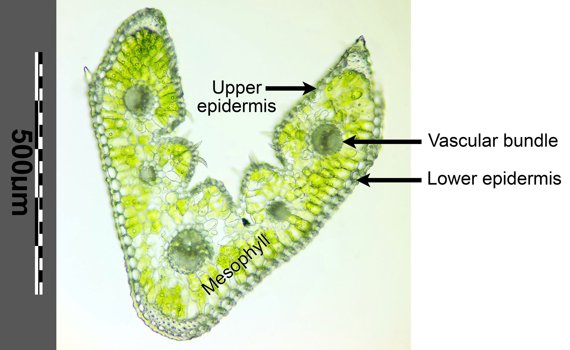
Fresh cross section of a leaf of furrowed fescue (Festuca rupicola) showing five veins, each surrounded by an inconspicuous bundle sheath made up of a ring of cells. Mesophyll cells are distributed evenly around the vascular bundles in this C3 plant. Photo by Stefan.Iefnaer (Wikimedia Commons, Creative Commons Attribution-ShareAlike 4.0 International license, image labeled, cropped, and resized).
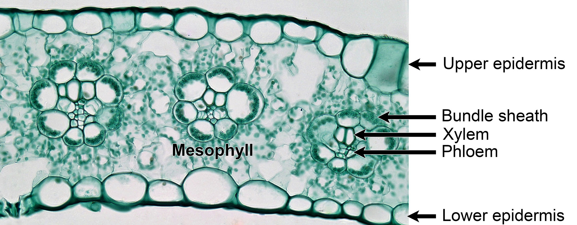
Fixed and stained cross section of a leaf of maize (corn, Zea mays) showing three veins, each with vascular tissue surrounded by a bundle sheath. Mesophyll cells encircle the bundle sheaths. Photo by Berkshire Community College (flickr, CC0 1.0 Univerisal/public domain dedication).
Reproductive structures
Inflorescences, spikelets, and florets
An inflorescence (group of flowers) in grasses may have a stalk, called a peduncle, to which branches are attached. It may also have a central axis, the rachis. The peduncle or rachis often bears branches on which reproductive units called spikelets are arranged.
Each spikelet bears one, several, or many florets. The florets may or may not be attached to a rachilla (central axis or stem of a spikelet). Typically, two bracts (small modified leaves) called glumes occur at the base of each spikelet, below the florets. The glumes protect the florets in the spikelet before they are mature. Spikelets may be sessile (lack a stalk) or pedicellate (have a pedicel, or stalk).
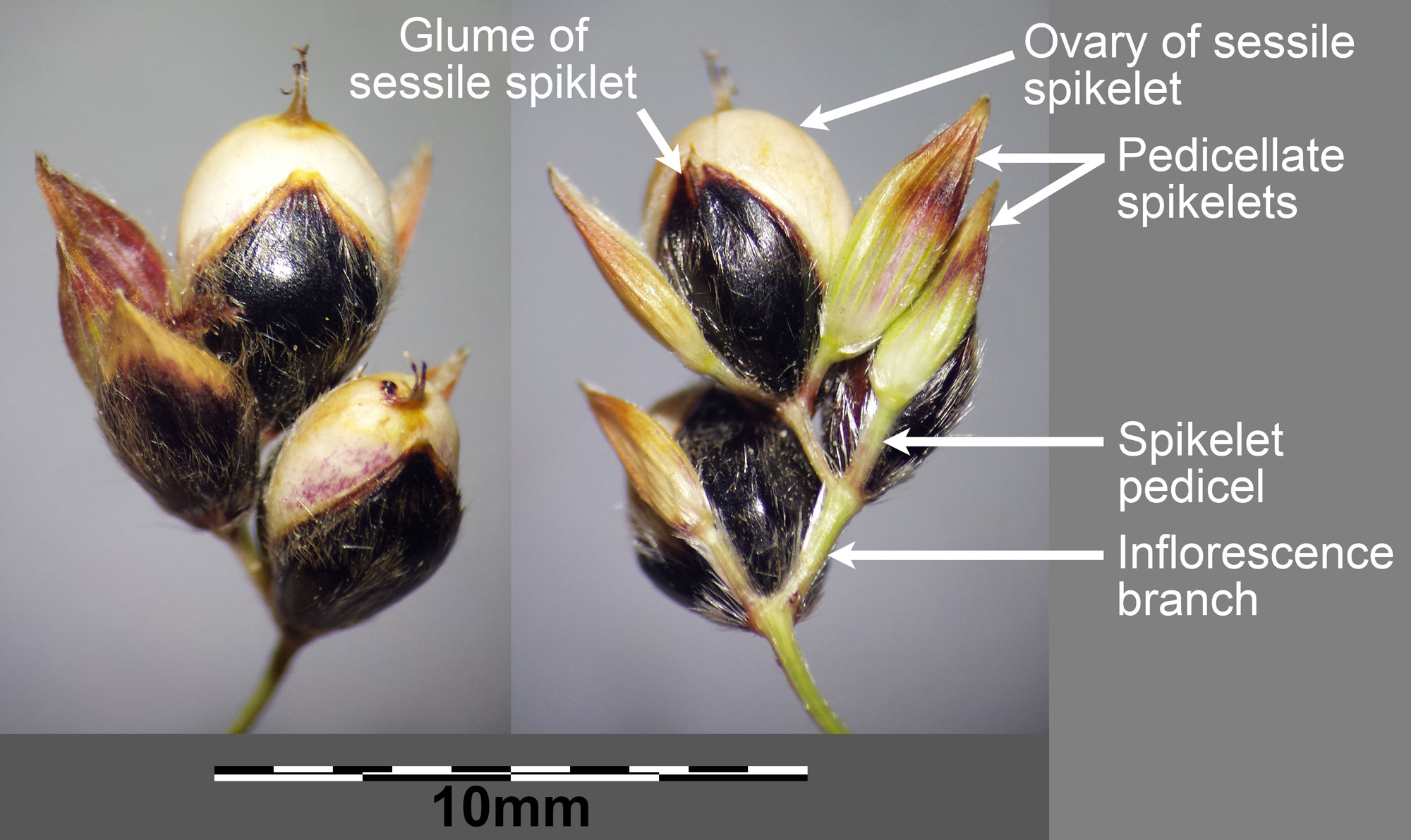
Sorghum (Sorghum bicolor) spikelets in two views. Sorghum has sessile and pedicellate spikelets. The sessile spikelets are bisexual (male and female), whereas the pedicellate spikelets may be male or sterile. Photo by Stefan.Iefnaer (Wikimedia Commons, Creative Commons Attribution-ShareAlike 4.0 International license, image resized and labeled).
The florets are usually attached to the rachilla in an alternate and distichous arrangement; in other words, the florets are attached one at a time to the rachilla and are arranged in two vertical rows. Often, the rachilla has a zig-zag course, changing direction at the attachment point of each spikelet.
Each floret consists of bracts and reproductive structures. Two types of bracts are attached at the base of each floret. The lower bract is the lemma, and the upper bract is the palea. The lemma and palea may be large enough to mostly or entirely cover the reproductive parts, although in some cases they are very small.
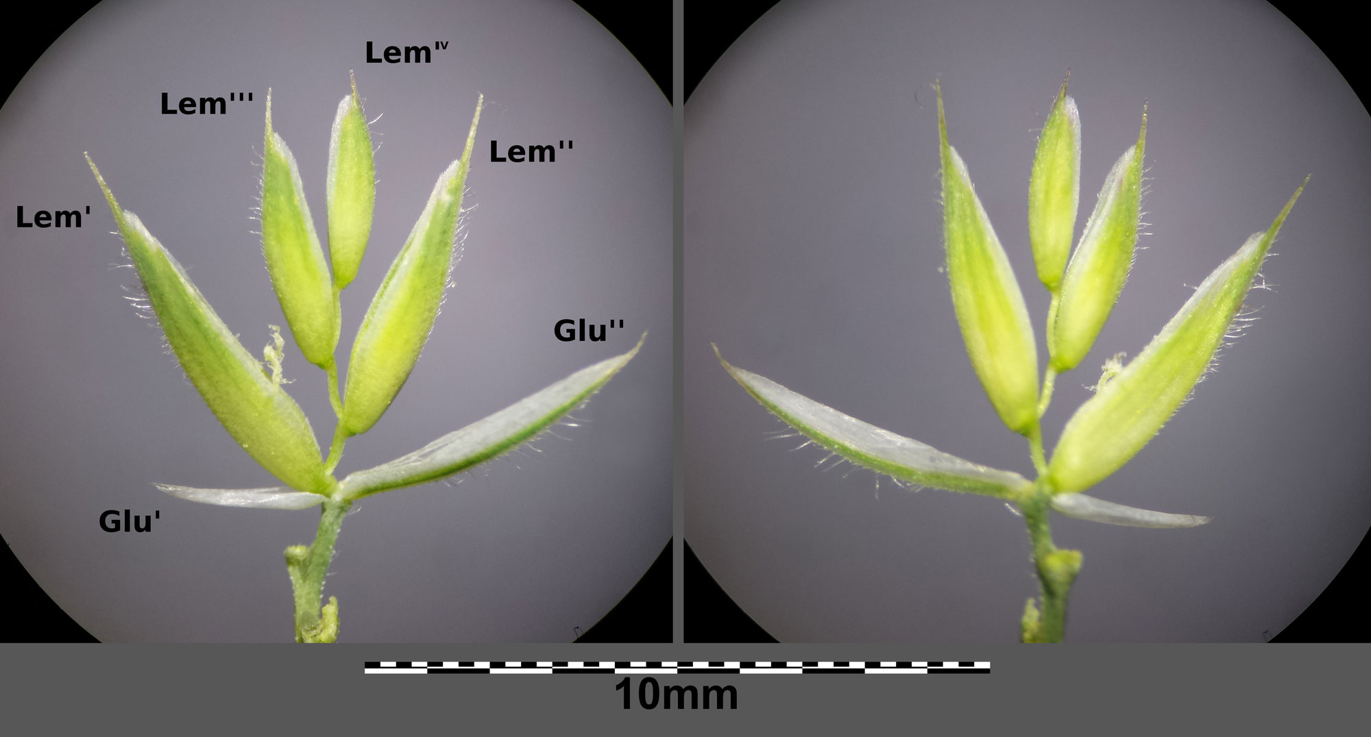
Cat grass (Dactylis glomerata subsp. glomerata) spikelet with four florets. GluI is the lower glume, GluII is the higher glume, and LemI to LemIX are the lemmas (lowest bracts) of the four florets attached to the rachilla above the glumes. This spikelet has been opened up so that its structure can be seen. Photo by Stefan.Iefnaer (Wikimedia Commons, Creative Commons Attribution-ShareAlike 4.0 International license, image resized).
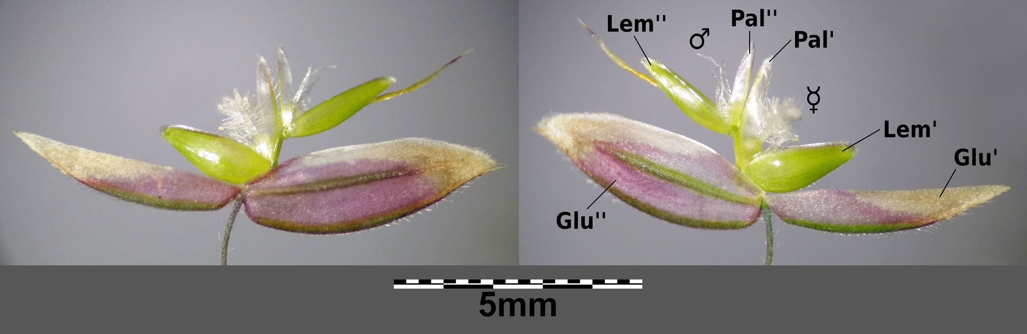
Velvet grass (Holcus lanatus) spikelet with two florets. GluI is the lower glume, GluII is the higher glume. Each of the two florets as a lemma (Lem) and palea (Pal). The lower flower is bisexual (has male and female parts) and the upper flower is male. This spikelet has been opened up so that its structure can be seen. Photo by Stefan.Iefnaer (Wikimedia Commons, Creative Commons Attribution-ShareAlike 4.0 International license, image resized).
The reproductive parts of the floret are attached above the lemma and palea. The reproductive parts may consist of stamens (pollen-producing male organs), a pistil (ovule-producing female organ), or both, with the stamens attached below the pistil. Florets that produce only pollen or ovules are unisexual; pollen-producing florets are called staminate or male florets and ovule-producing florets are called carpellate, pistillate, or female florets. Florets with both stamens and pistils are bisexual. Sometimes, florets may be sterile, which means that they do not have functional reproductive parts.
Florets that produce pollen (male and bisexual florets) most commonly have three stamens. Each stamen consists of a filament (stalk) and an anther (pollen-producing structure, usually with four fused pollen sacs).
Florets that produce ovules (female and bisexual florets) have one pistil, consisting of an ovary (ovule-containing structure) with two or three feathery stigmas (structures that receive pollen). The stigmas may be elevated on stalks known as styles. The ovary contains a single ovule, which is an immature seed.
Lodicules (small bracts, which may look like swellings or bumps) are typically found below the reproductive structures. The lodicules enlarge to help the floret open.
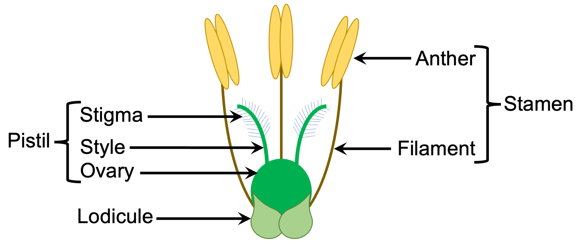
Diagram of a bisexual grass floret with lemma and palea removed. Diagram by Elizabeth J. Hermsen (Evolution/Earth@Home).
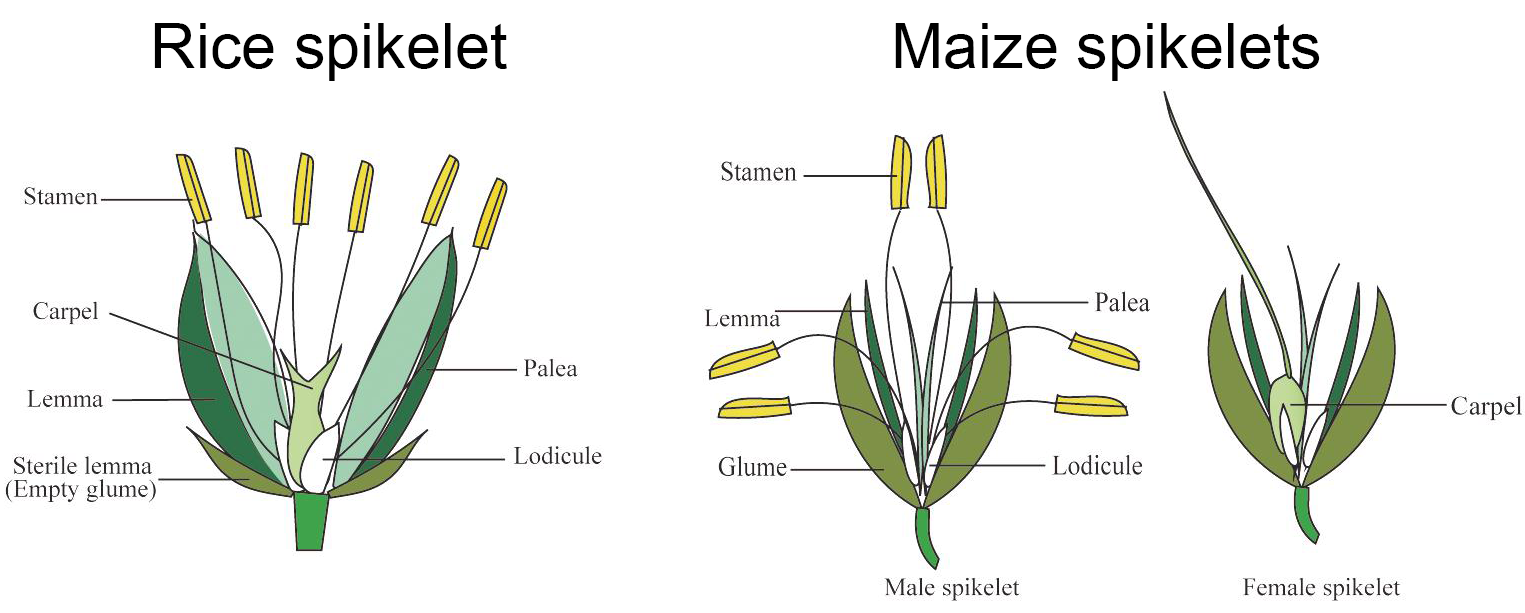
Diagram of a bisexual rice spikelet with one floret and diagrams of maize spikelets, a male spikelet with two florets and a female spikelet with one fertile floret. The structure labeled "carpel" is the pistil. Source: Modified from Figure 1 in Shen et al. (2021) Frontiers in Plant Science 12:635500 (Creative Commons Attribution 4.0 International license).
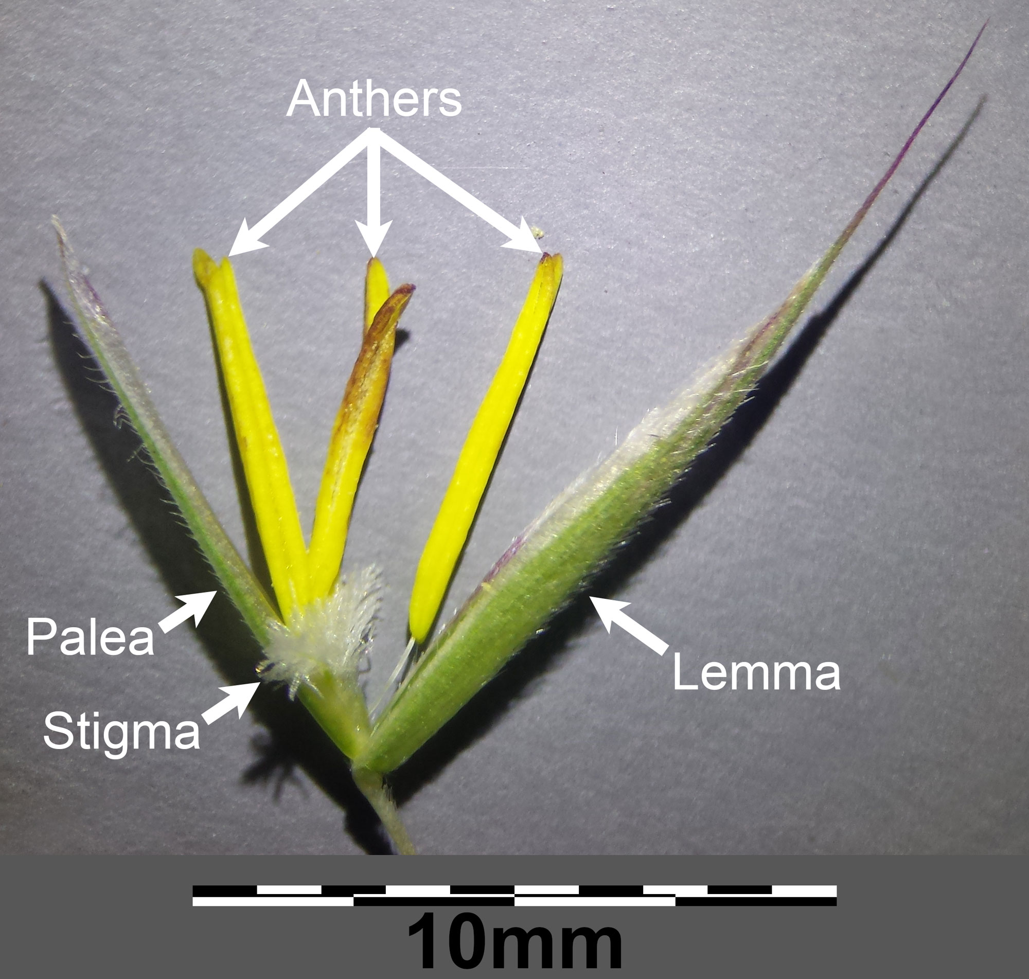
Bisexual foret of erect brome (Bromus erectus) showing the lemma, palea, and reproductive structures. The lodicules cannot be seen in this photo. Photo by Stefan.Iefnaer (Wikimedia Commons, Creative Commons Attribution-ShareAlike 4.0 International license, image resized and labeled).
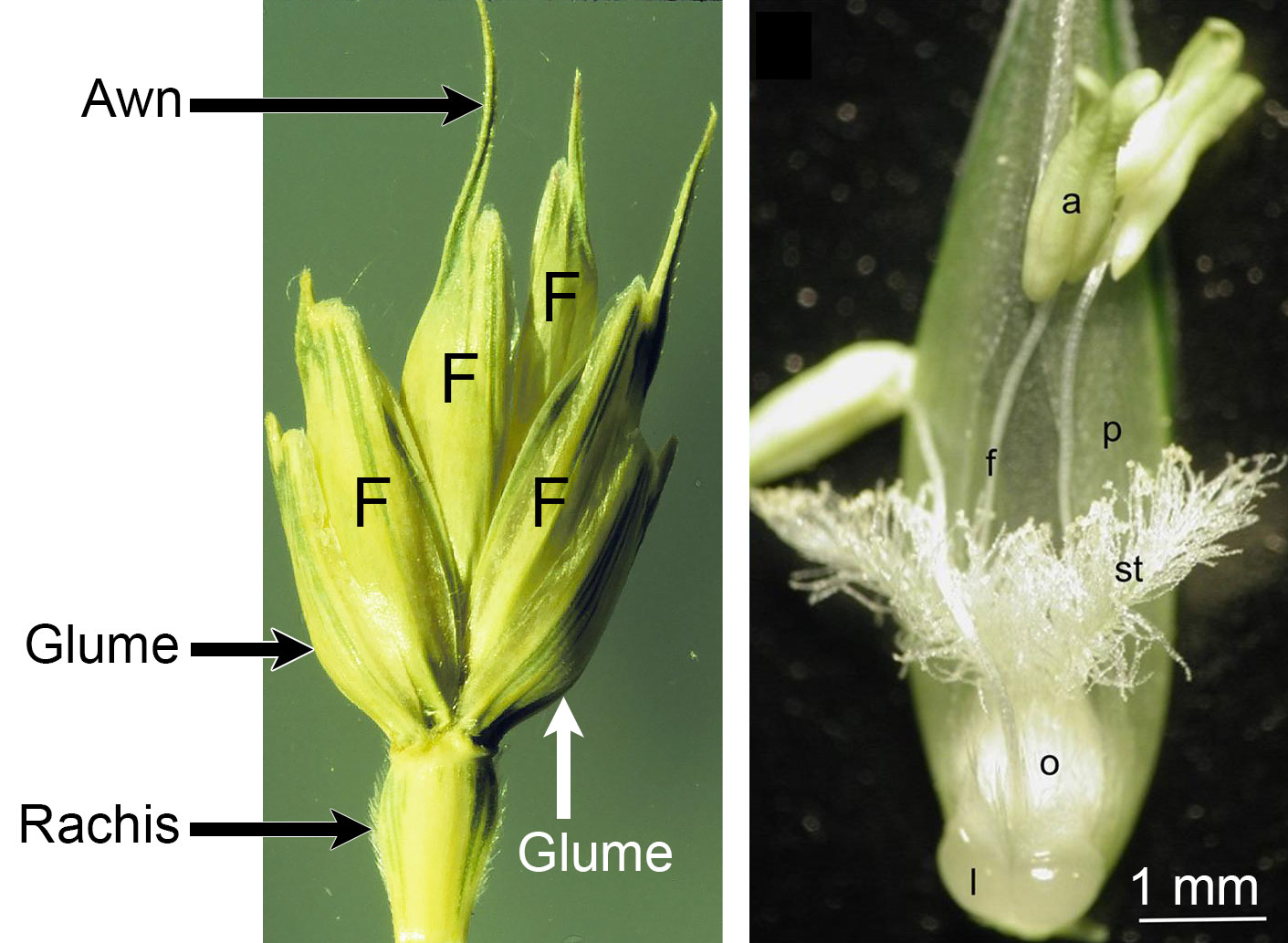
Wheat (Triticum aestivum) spikelet and floret. Left: Spikelet of wheat showing part of the rachis (inflorescence axis), the two glumes, and four florets (each marked F). An awn is an a pointed extension; in this wheat spiklet, the long awns are extensions of the lemmas of the florets. Right: Part of a single floret dissected to show the reproductive structures. a = anther, f = filament, l = lodicule, o = ovary, p = palea, st = stigma. Left photo by Matt Lavin (flickr, Creative Commons Attribution-ShareAlike 2.0 Generic license, image cropped, resized, labels added); right photo from Fábián et al. (2019) Frontiers in Plant Science (Creative Commons Attribution 4.0 International license, figure number obscured, label added to scale bar).
Fruits
After pollination and fertilization, female and bisexual flowers will mature to produce fruits. The grass fruit is the caryopsis or grain. A grain is fruit that contains one seed. The thin fruit wall is fused to the thin seed coat. The seed contains a tiny grass embryo and endosperm, which is stored food for the young grass.
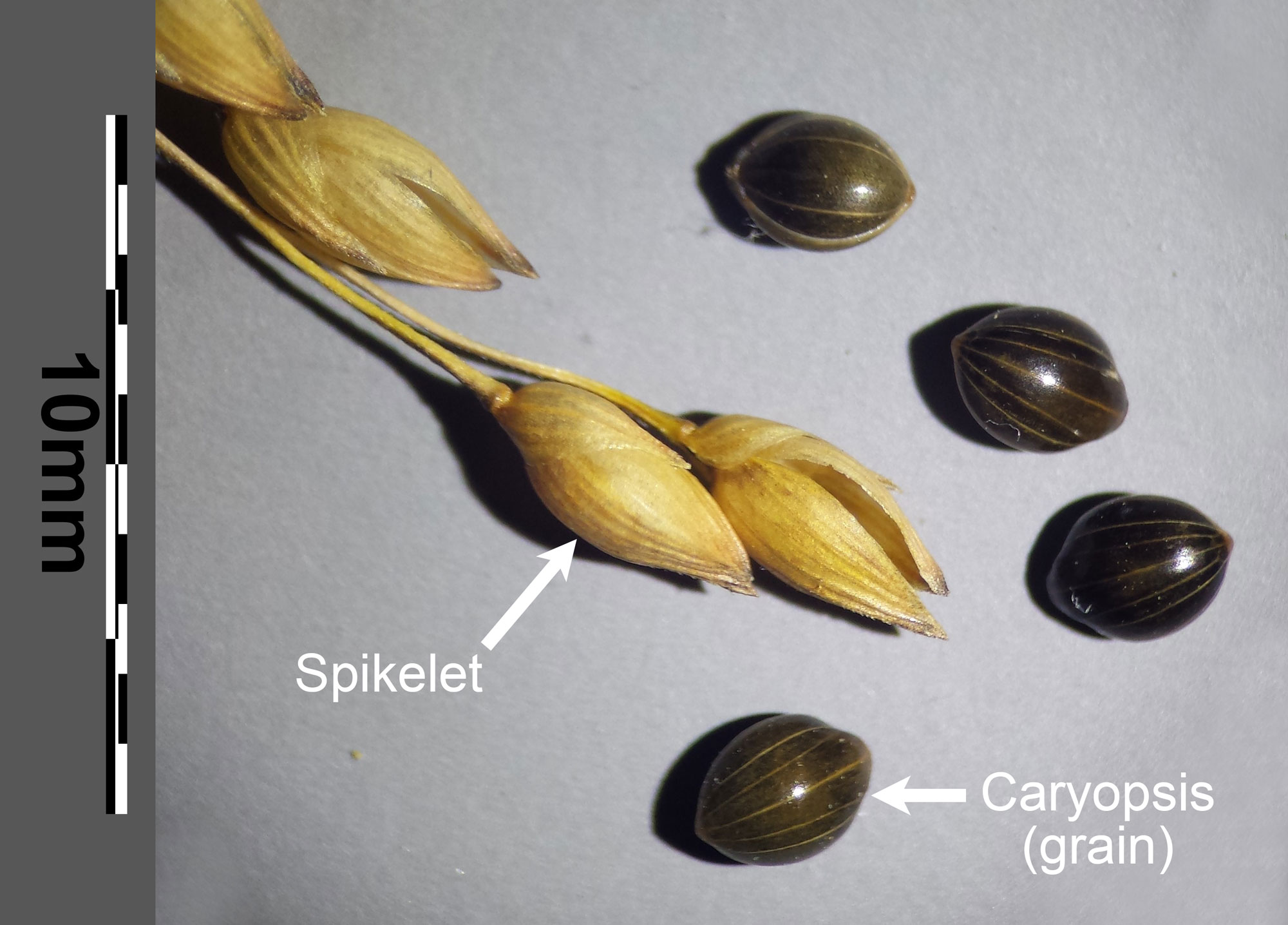
Spikelets and caryopses (grains) of broomcorn millet (Panicum milliaceum subspecies agrcola). Photo by Stefan.Iefnaer (Wikimedia Commons, Creative Commons Attribution-ShareAlike 4.0 International license, image resized and labeled).
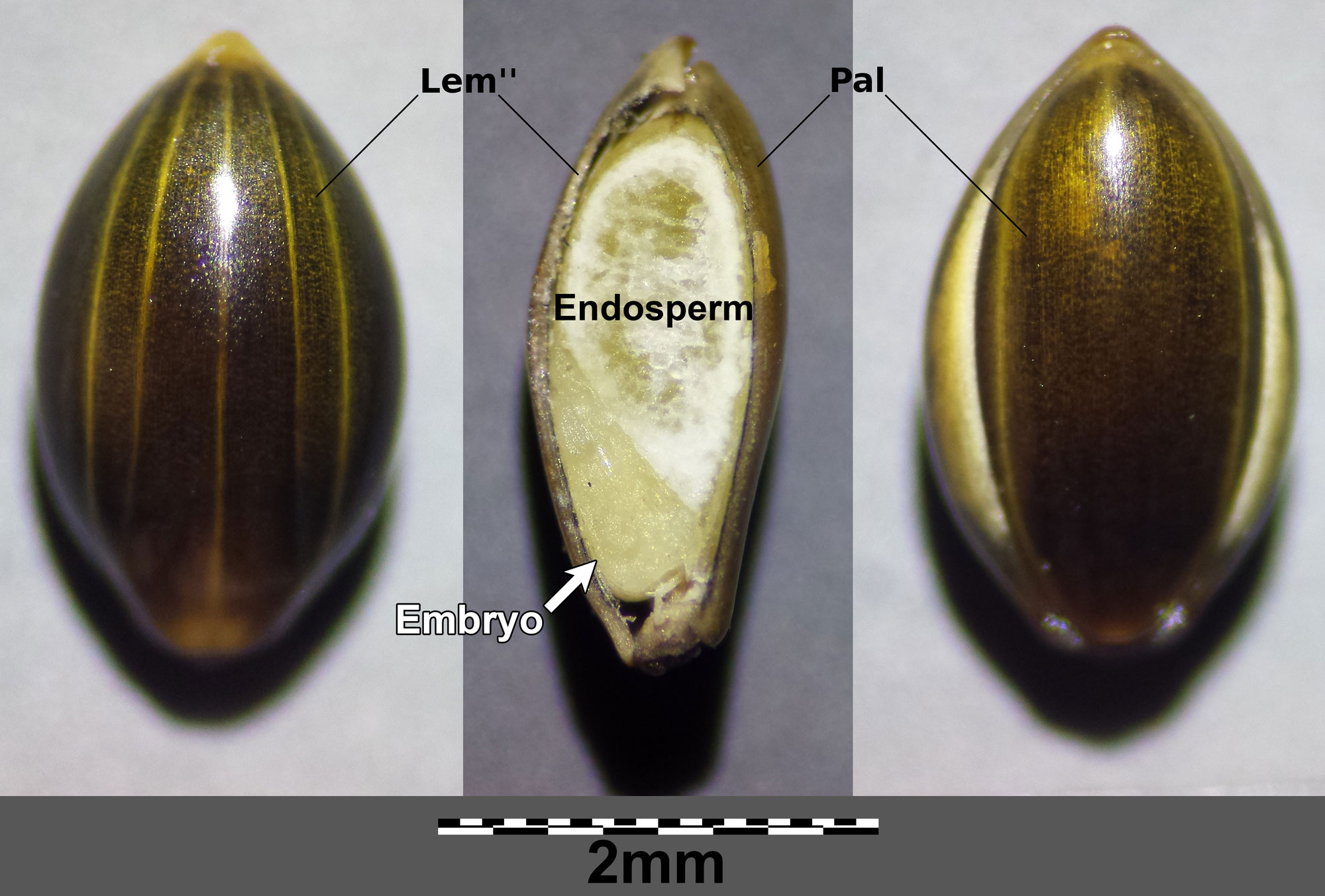
Caryopsis (grain) of broomcorn millet (Panicum milliaceum subspecies ruderale) in three views. In this caryopsis, the lemma and palea form part fo the covering of the fruit. Lem = lemma, Pal = palea. Left: Outer surface showing lemma. Center: Longitudinal section of fruit showing embryo (young grass) and endosperm (food for the embryo). Right: Outer surface showing palea. Photo by Stefan.Iefnaer (Wikimedia Commons, Creative Commons Attribution-ShareAlike 4.0 International license, image resized and some labels added).
Resources
Additional resources from the Paleontological Research Institution
Digital Encyclopedia of Ancient Life: Introduction to Vascular Plant Structure: https://www.digitalatlasofancientlife.org/learn/embryophytes/tracheophytes/
Digital Encyclopedia of Ancient Life: Angiosperms (Flowering Plants): https://www.digitalatlasofancientlife.org/learn/embryophytes/angiosperms/
Other websites
Glossary of botanical terms used in Poaceae (Flora of China online): http://flora.huh.harvard.edu/floradata/002/vol22/poaceaeglossary.htm
Poaceae (Graminae) (University of Hawai'i): http://www.botany.hawaii.edu/faculty/carr/po.htm
Scientific articles and books
Esau, K. 1977. Anatomy of seed plants, 2nd. ed. John Wiley and Sons, New York.
Evert, R. F., and S. E. Eichhorn. 2013. Raven Biology of Plants, 8th ed. W.H. Freeman and Company Publishers, New York, New York.
Kellogg, E. A. 2015. Flowering Plants, Monocots, Poaceae. The Families and Genera of Vascular Plants 13, 416 pp. (K. Kubitzki, ed.). Springer International Publishing, Switzerland. https://doi.org/10.1007/978-3-319-15332-2_1
Palmer, P. G., and A. E. Tucker. 1981. A scanning electron microscope survey of the epidermis of East African Grasses, I. Smithsonian Contributions to Botany 49, 84 pp. https://doi.org/10.5962/bhl.title.131623
Rao, X., and R. A. Dixon. 2016. The differences between NAD-ME and NADP-ME subtypes of C4 photosynthesis: More than decarboxylating enzymes. Frontiers in Plant Science 7:1525. https://doi.org/10.3389/fpls.2016.01525
Twiss, P. C., E. Suess, and R. M. Smith. 1969. Morphological classification of grass phytoliths. Proceedings of the Soil Science Society of America 33: 109-115.