

Page snapshot: Introduction to the structure of the maize plant, including the morphology (form) and anatomy (internal structure) of stems, roots, and leaves. Also covers the organization of the reproductive structures.
Topics covered on this page: Introduction; Stems; Roots; Leaves; Leaf morphology; Leaf anatomy; Reproductive structures; Male (staminate) structures; Female (pistillate or carpellate) structures; Fruits; Resources.
Credits: Funded by the National Science Foundation. Any opinions, findings, and conclusions or recommendations expressed in this material are those of the author(s) and do not necessarily reflect the views of the National Science Foundation. This page incorporates content prepared for a revised manuscript of the Teacher-Friendly Guide to the Evolution Maize (Carlyn Buckler, Dhyan Palanichamy, and Andrielle Swaby, 2019). Additional content and revisions by Elizabeth J. Hermsen (2022).
Updates: Page last updated January 11, 2023.
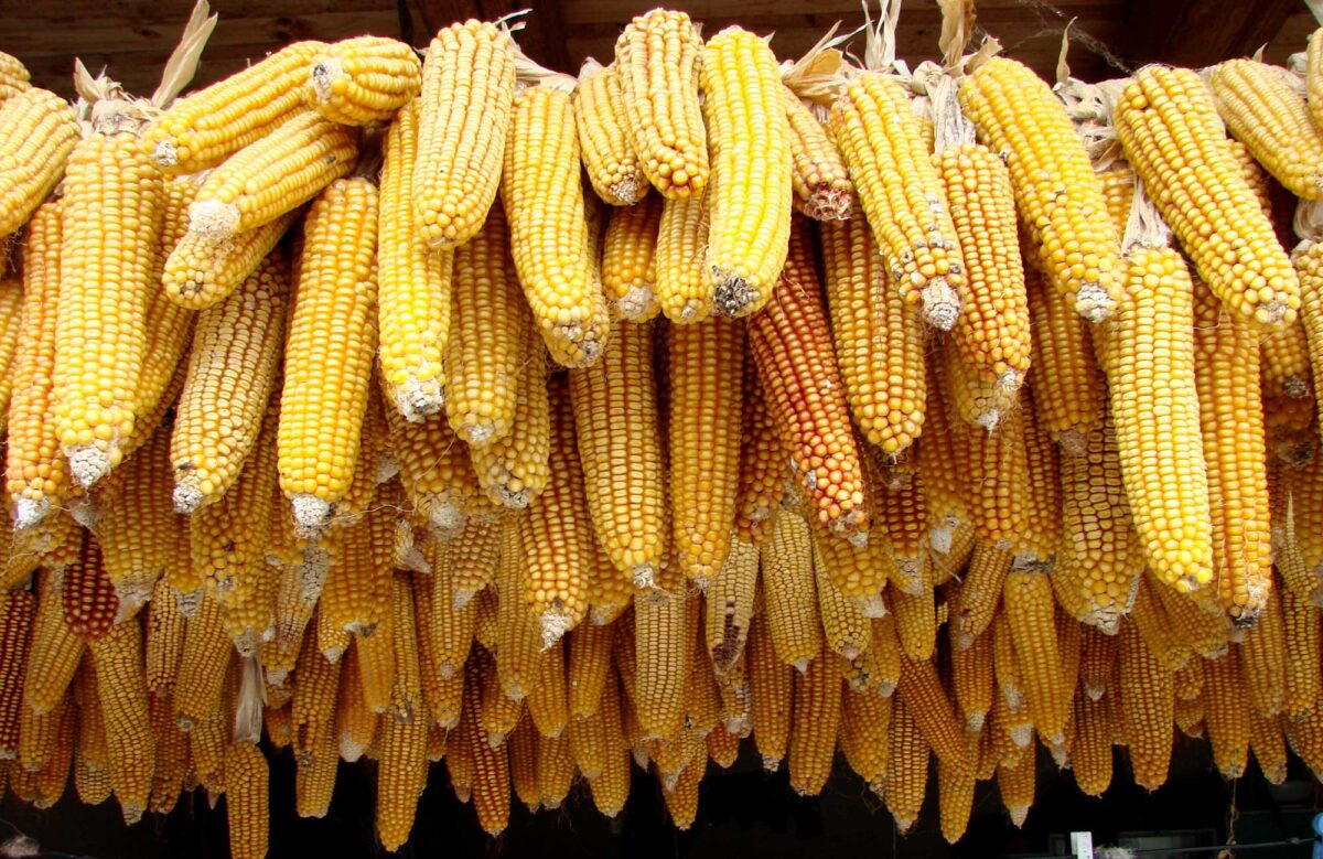
Image above: Ears of corn, Romania. Photo by Adam Jones/Adam 63 (Wikimedia Commons, Creative Commons Attribution-ShareAlike 3.0 Unported license, image cropped and resized).
Introduction
The purpose of this page is to introduce the basic structure of the maize plant. Generally speaking, maize plants are organized like other grasses in tribe Andropogoneae (the Bluestem or Sorghum Tribe), except that they have been modified by the process of domestication. The domestication of maize altered it from its wild form by reducing the amount of stem branching and by radically modifying its female reproductive structures, called ears. Maize ears are unusually large with many rows of soft-walled kernels (fruits) that stay attached to the ear at maturity.
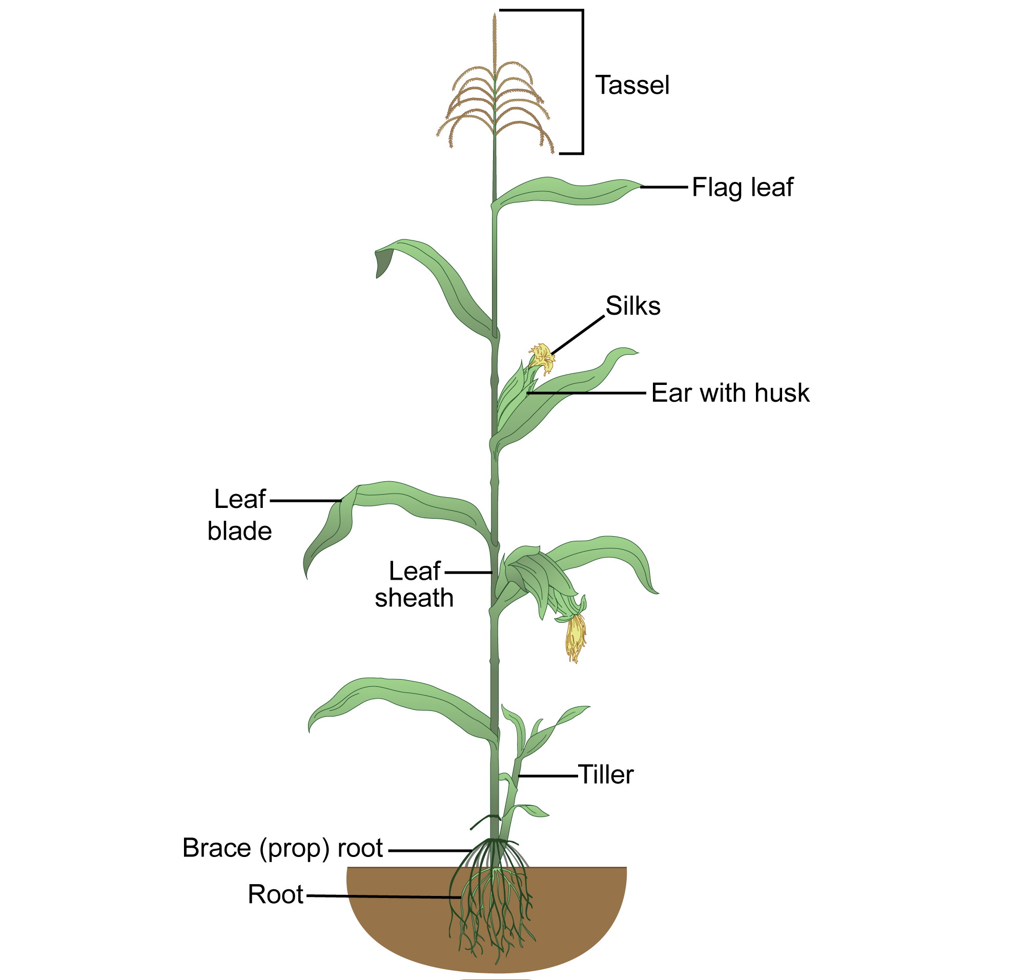
Diagram of a mature maize (Zea mays) plant. Diagram by LadyofHats (Wikimedia Commons, CC0 1.0 Universal/public domain dedication, image relabeled).
For background on the general structure of the grass plant, visit the following page:
For background on the structure of grasses in Tribe Andropogoneae, visit the following page:
Stems
The stem of a maize plant is a long central stalk that supports every part of the plant. The only branches on the stem are those that support the reproductive parts. The strength of the stem allows maize to grow very tall; maize plants can reach as much as 12 feet (4 meters) in height, depending on the variety. Only one leaf grows per node, and stalks can have 20 or more nodes.
Unlike in many other grasses, the interior of a maize stem is solid in both nodal and internodal regions. Vascular bundles are scattered throughout the stem when it is viewed in cross section.
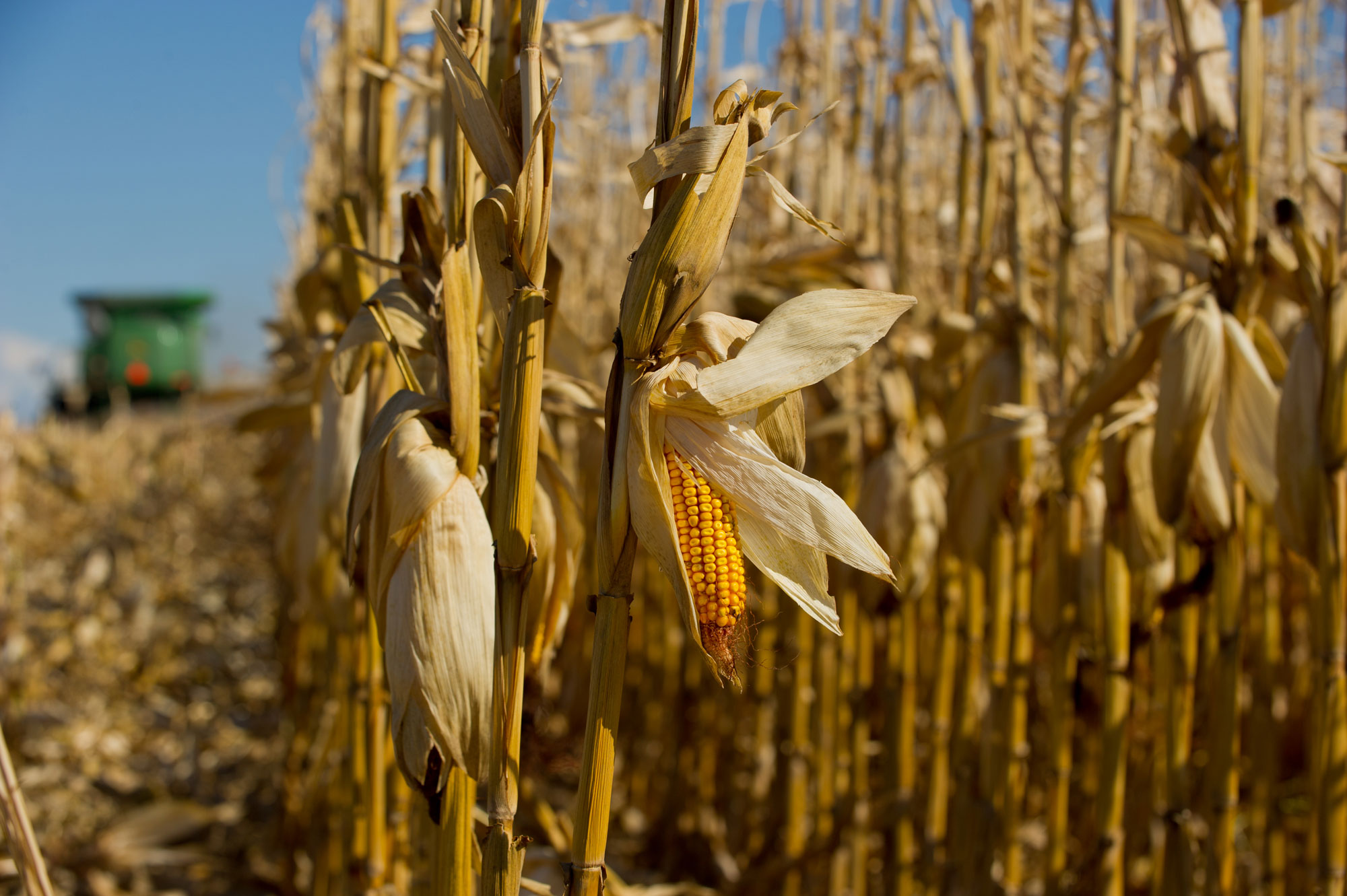
Corn (Zea mays) dry cornstalks during harvest. Photo by United Soybean Board (flickr, Creative Commons Attribution 2.0 Generic license, image resized).

Cross-section of a maize (Zea mays) stem. The vascular bundles (water- and food-conducting tissue) are scattered throughout ground tissue in the stem. Photo by John Houseman and Matthew Ford (Wikimedia Commons, Creative Commons Attribution-ShareAlike 4.0 International license, image modified from original).
Roots
Maize plants have three types of roots: the radicle or primary root, the seminal roots, and the nodal roots.
The radicle (called the primary root once it emerges from the kernel) makes up the lower part of the maize embryo. Seminal roots ("seed roots") are roots that first start to form on the embryo above the radicle. The primary root is the first root to emerge from the corn kernel when the seedling starts to grow. Three to four seminal roots appear after the radicle has emerged. The primary root and seminal roots are responsible for absorbing water and nutrients in order to establish the young seedling.
The radicle and the seminal roots will eventually die, and their function will be taken over by nodal roots. Nodal roots are adventitious roots, or roots that grows from the stem. Nodal roots arise from the stem at the nodes, places where leaves and their associated lateral buds develop. On a healthy maize plant, nodal roots can grow up to six feet (two meters) deep underground.
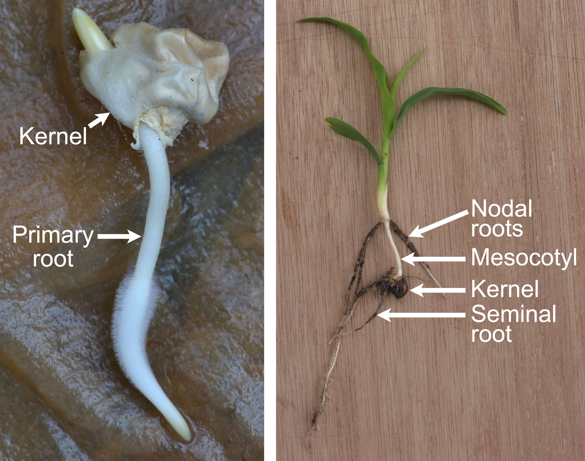
Germinated kernels (grains or caryopses) of maize (Zea mays). Left: A germinated kernel with a primary root bearing root hairs protruding from its lower side; a bit of a the coleoptile (shoot sheath) is extending from its upper side. Right: Seedling of maize bearing seminal roots and first nodal roots separated by an extended mesocotyl (a transition region between the primary root and the aboveground parts of the plant). The first nodal roots will remain completely underground. Left photo and right photo by Rasbak (Wikimedia Commons, Creative Commons Attribution-ShareAlike 3.0 Unported license, images cropped, left image resized, labels added).
Special nodal roots, called brace roots or prop roots, form at nodes above the soil surface. These roots function primarily to support the aboveground parts of the plant once it grows tall. When brace roots reach the soil, they can also perform regular root functions such as water and nutrient uptake. Over the course of a maize plant’s life, as many as 70 nodal roots will arise from the stem, both above and below the ground.
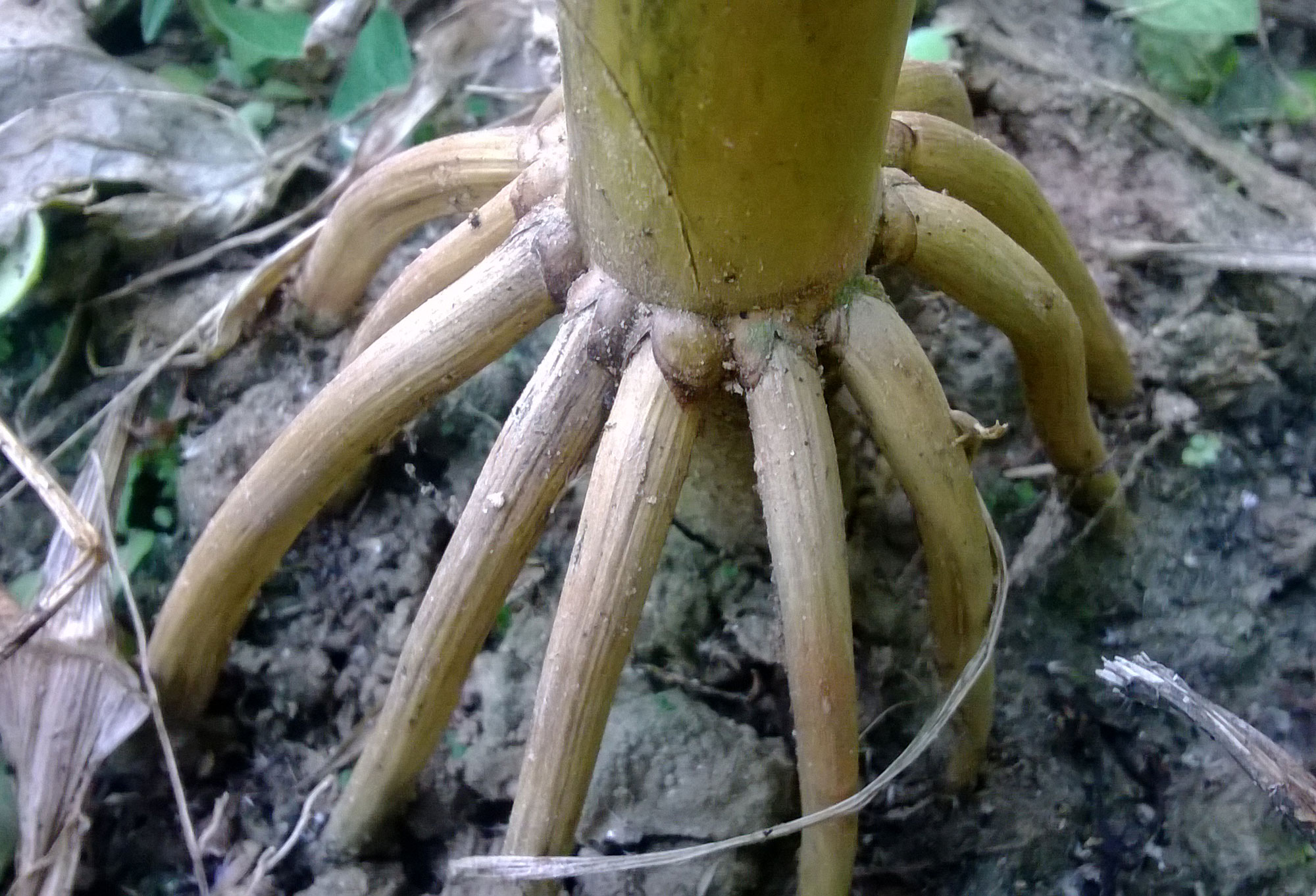
Prop roots of maize (Zea mays). Photo by Krish Dulal (Wikimedia Commons, Creative Commons Attribution-ShareAlike 3.0 Unported license, image cropped and resized).
Leaves
Leaf morphology
As in other grasses, maize leaves alternate along the maize stem. This arrangement spreads the leaves out to maximize the amount of light they can capture.
Maize leaves are organized like the leaves of other grasses. Each leaf consists of a sheath that wraps around the stem and an elongated blade that sticks out from the side of the stem and intercepts sunlight. The place where the sheath and the blade come together is the collar. The major leaf veins are parallel.
Each new leaf begins to grow from a node on the inside of the older (lower) adjacent leaf’s sheath. The last foliage leaf on the stem (the highest leaf) is called the flag leaf. The flag leaf is the leaf directly beneath the tassel.
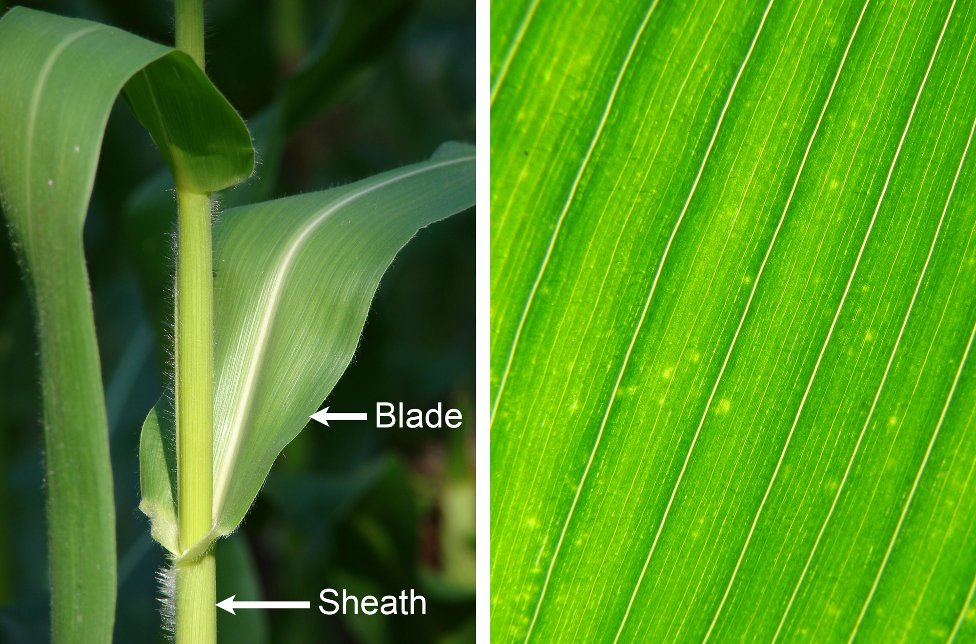
Maize (Zea mays) foliage leaves. Left: Leaves on a stem, showing sheaths and blades. Right: Close-up of a leaf blade showing parallel venation. Left photo (Creative Commons Attribution 3.0 Unported license) and right photo (Creative Commons Attribution 2.0 Generic license) by Forest & Kim Starr (Wikimedia Commons, images cropped and resized).
Leaf anatomy
Leaf epidermis
The epidermis of the maize leaf is similar to that of other grasses. It has elongated long cells and shorter short cells that may contain silica (silica cells) or suberin (cork cells). The long cells have wavy side walls. The silica cells have crossed-shaped silica bodies (phytoliths).
Stomata (pores) occur between some of the long cells. The stomata are opened and closed by dumbbell-shaped guard cells, which are flanked on either side by triangular subsidiary cells.
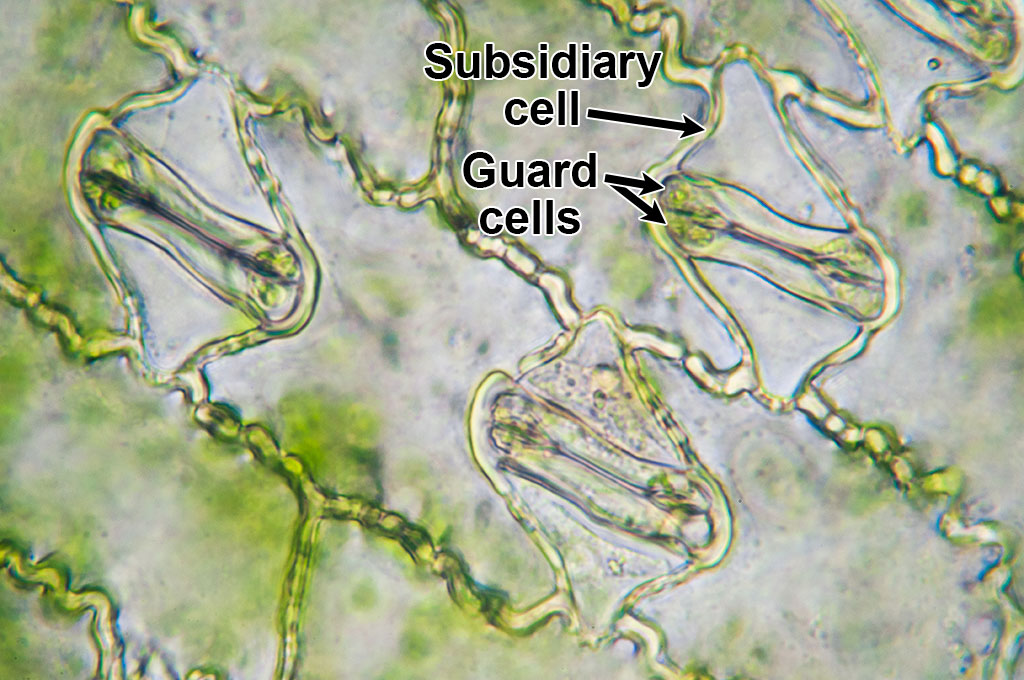
Microscopic image of maize (corn, Zea mays) epidermis showing three stomata with guard cells and subsidiary cells. In this image, the stomata are closed. The cells between the stomata are interstomatal cells, a type of long cell (although in this image, these cells are relatively short). Photo by Umberto Salvagnin (flickr, Creative Commons Attribution 2.0 Generic license, image labeled).
Leaf vascular bundles and mesophyll
Like other members of Tribe Andropogoneae, maize is a C4 plant. Each vascular bundle has a bundle sheath made up of one layer of cells with large chloroplasts near their outer walls. As is common in C4 plants, mesophyll cells are arranged concentrically around the bundle sheath.
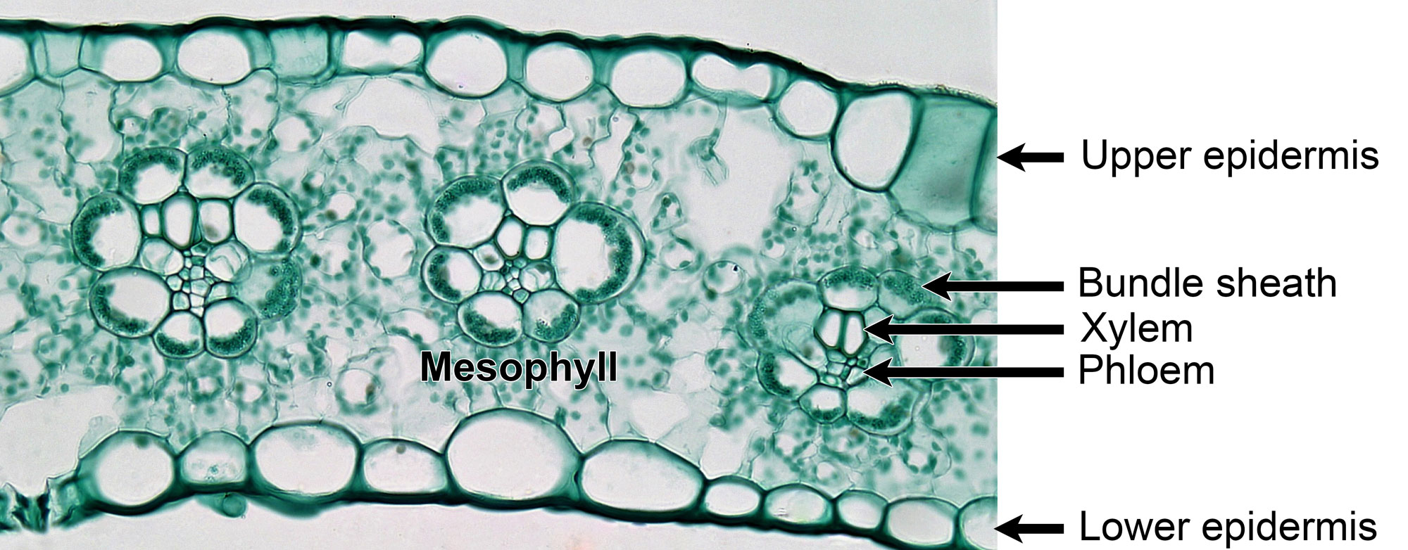
Fixed and stained cross section of a leaf of maize (corn, Zea mays) showing three veins, each with vascular tissue surrounded by a bundle sheath. Mesophyll cells encircle the bundle sheaths. Photo by Berkshire Community College (flickr, CC0 1.0 Univerisal/public domain dedication).
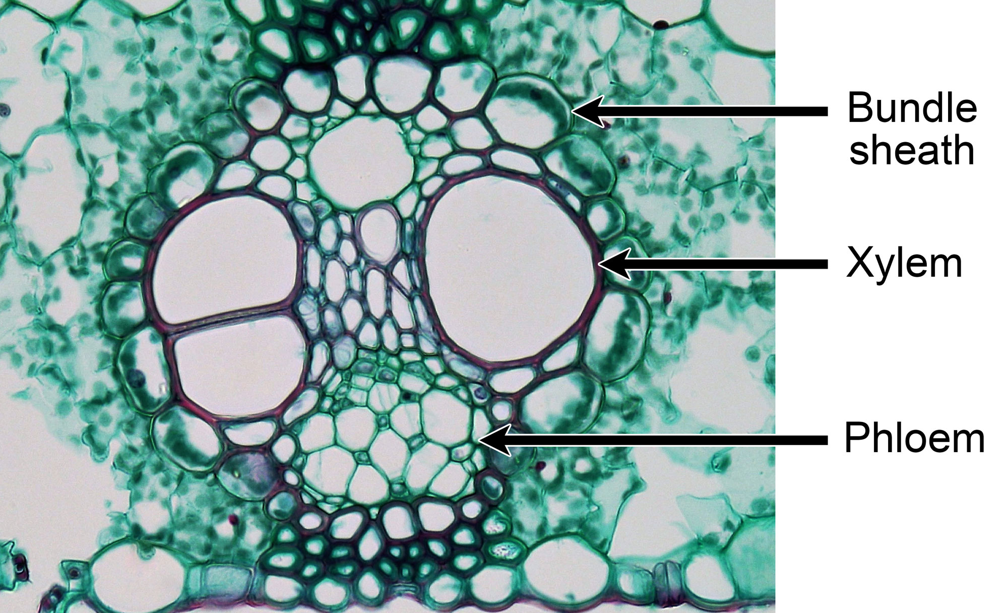
Fixed and stained cross section of a leaf of maize (corn, Zea mays) showing a large vascular bundle encircled by a single-layered bundle sheath with cells containing chloroplasts. Maize is a C4 grass. Photo by Berkshire Community College (flickr, CC0 1.0 Univerisal/public domain dedication).
Reproductive structures
Maize plants are monoecious, meaning that flowers are unisexual (male or female), but both male (staminate, or pollen-producing) and female (pistillate, or ovule-producing) flowers grow on the same plant.
Male (staminate) structures
The male flowers are grouped in an inflorescence called a tassel at the tip of the stem. Maize tassels consist of a central stem bearing branches (also called rames) on its sides. Each branch bears many pairs of spikelets. Each spikelet pair includes one pedicellate (stalked) spikelet and one sessile (stalkless) spikelet.
Each spikelet has two male florets (tiny flowers). Each male floret has three stamens consisting of a stalk-like filament and an anther, or pollen-producing structure. Depending on the variety of maize, the anthers can vary in color between purple, red, and yellow.
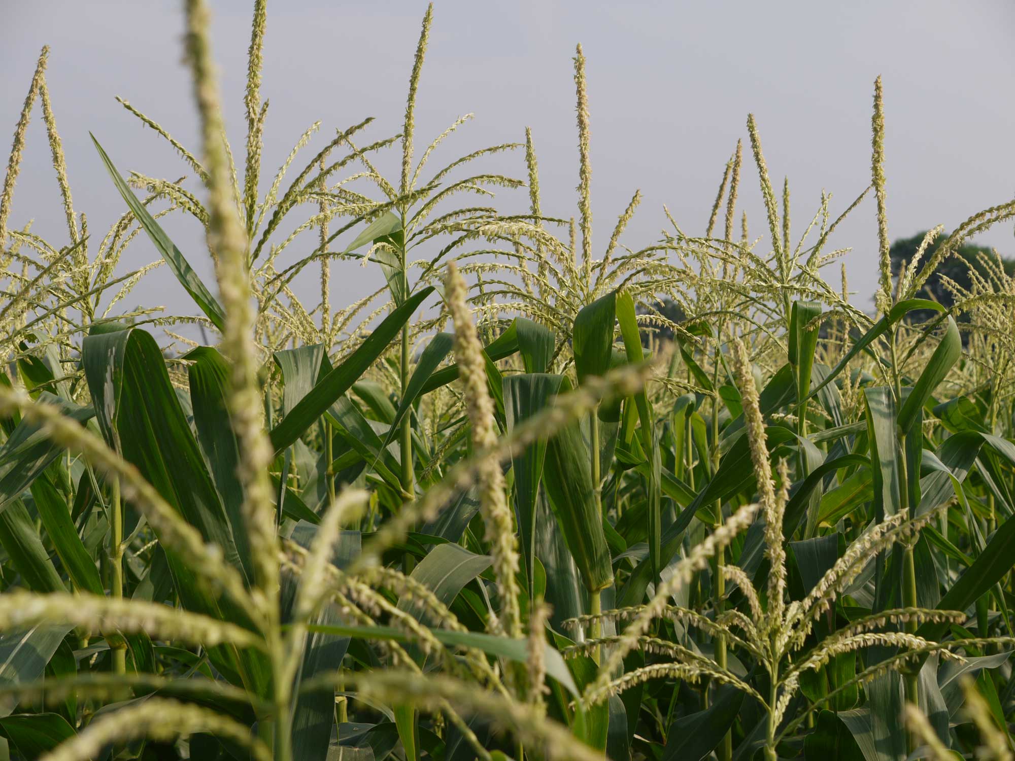
Tassels of maize (Zea mays). Photo by Dinesh Valke (flickr, Creative Commons Attribution-ShareAlike 2.0 Generic license, image resized).
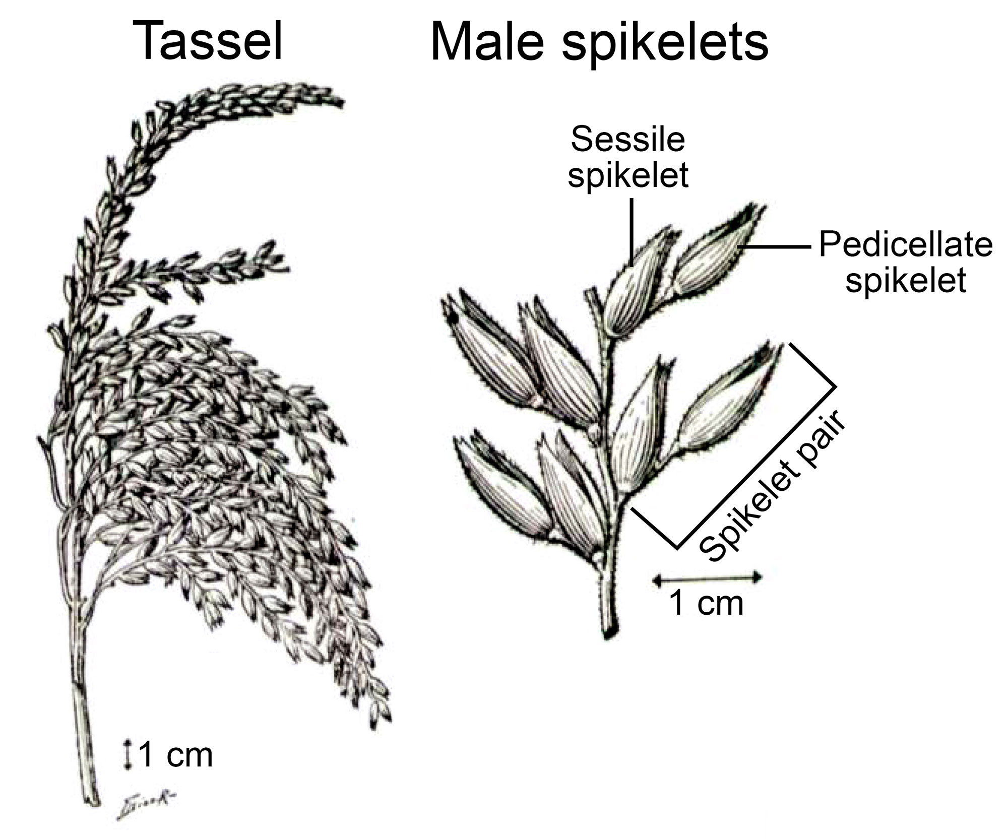
Tassel and male (staminate) spikelets of maize. Left: A tassel. Right: Detail of part of one of the tassel branches showing pairs of male (staminate) spikelets attached to a branch. Source: From figure 1 in Anderson and Brown (1948) Annals of the Missouri Botanical Garden 35: 323-336 (Biodiversity Heritage Library, Creative Commons Attribution-NonCommercial-ShareAlike 3.0 Unported license, image resized, labeled, contrast adjusted).
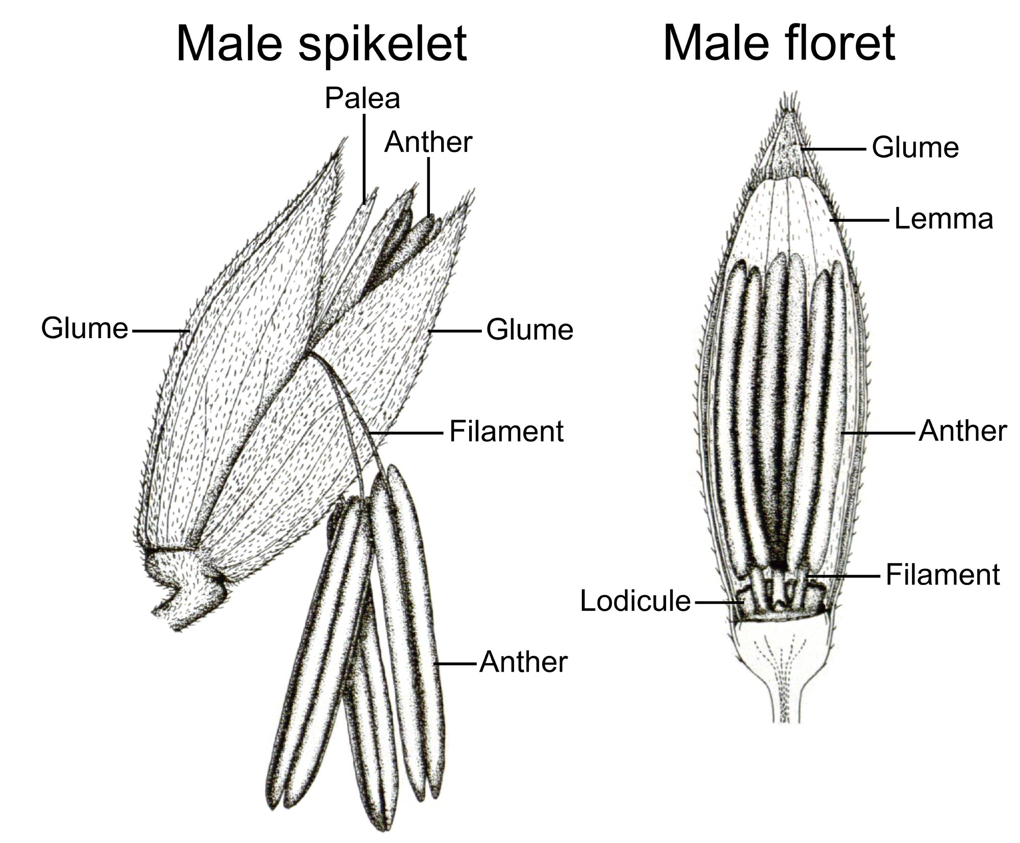
Diagrams of a maize male (staminate) spikelet and male (staminate) floret. Left: Male spikelet with two florets, one of which is open with stamens protruding. Note that the lemmas are not labeled because they are under the glumes, and thus not visible in the drawing. Right: A single dissected male floret. Note that the palea is not labeled because it has been removed to expose the stamens. Source: Plate 5, figs. 1 and 2 from Weatherwax (1916) Bulletin of the Torrey Botanical Club 43: 127-144 (Biodiversity Heritage Library, public domain).
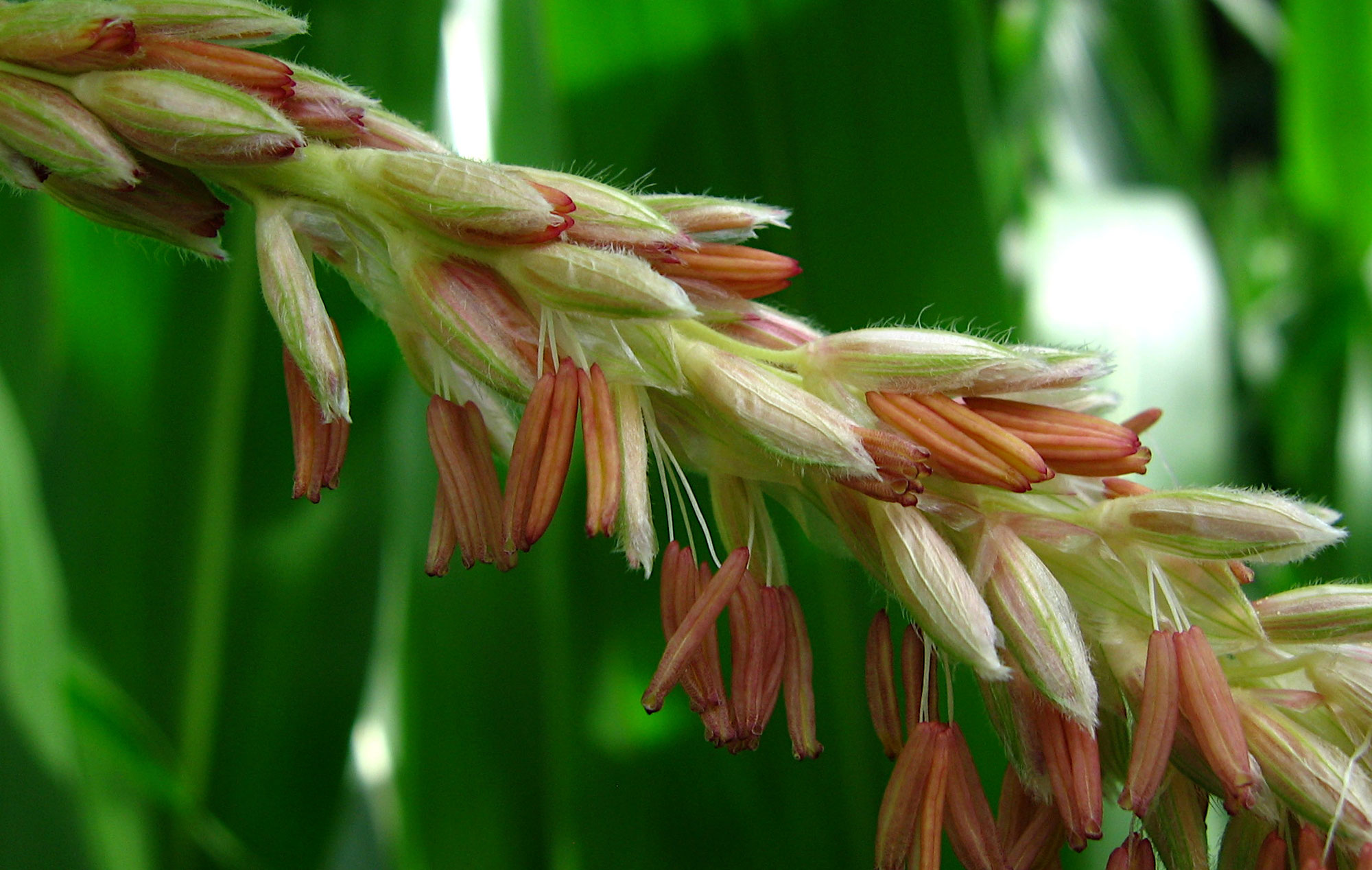
Male (staminate) spikelets of maize. Anthers are protruding from some of the spikelets. Source: Kostka Martin (Wikimedia Commons, CC01.0/public domain dedication).
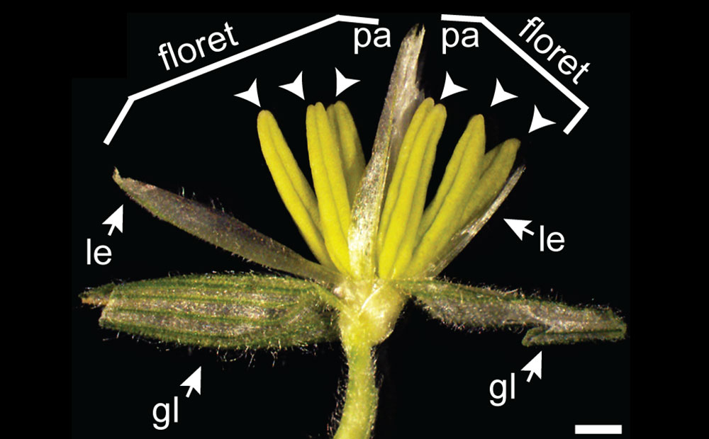
Male (staminate) spikelet of maize opened and labeled to show the parts of the two florets. Labels: gl = glume, le = lemma, pa= palea, and arrowheads = anthers. Scale bar = 2 millimeters. Source: From figure 2 in Field and Thompson (2016) PLoS ONE 11(1): e0146534 (Creative Commons Attribution 4.0 International license, image cropped and more background added).
Female (pistillate or carpellate) structures
Female maize flowers are arranged in ears (also called rames or spikes), which occur at the ends of short, thick branches on the sides of the main stem. A series of modified leaves surround each ear, forming a structure called a husk. The thick central stem of the ear is called a cob. The female spikelets are in pairs like the male spikelets. Each cob may have as many as 24 rows of spikelets.
Each spikelet has one fertile floret and one sterile floret. The sterile floret is made up of just a lemma and a palea. The fertile floret includes a lemma, a palea, and a pistil. Each fertile floret has one ovary and a short style with two long, fused stigmas, which are called a silk. The two glumes at the base of each female spikelet cover much of the ovary.
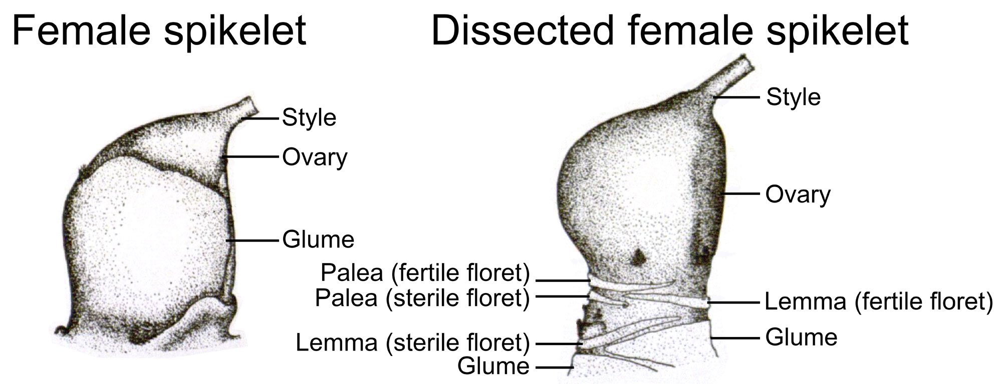
Diagrams of a maize female (pistillate or carpellate) spikelet. Left: Female spikelet with the bracts attached. Notice that the glumes cover much of the ovary. The style and stigma are very long, so they have been cut off in the image. Right: A dissected female spikelet. Note that the bracts (glumes, lemmas, and paleas) have been removed, leaving only their bases. The style and stigma are very long, so they have been cut off in the image. Source: Plate 5, figs. 3 and 4 from Weatherwax (1916) Bulletin of the Torrey Botanical Club 43: 127-144 (Biodiversity Heritage Library, public domain).
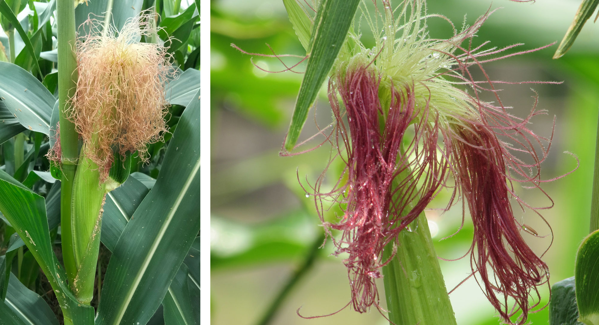
Maize silk, or stigmas. Left: Silks sticking out of the top of an ear of maize. Right: Red maize silks. Left photo by ALeL99 (Wikimedia Commons, Creative Commons Attribution-ShareAlike 4.0 International license), right photo by Bijay chaurasia (Wikimedia Commons, Creative Commons Attribution-ShareAlike 3.0 Unported license), images cropped and resized.
Fruits
Following pollination and fertilization, the fertile female florets mature into fruits. Maize fruits are often called kernels. Unlike in most andropogonoid grasses, ears of corn do not disarticulate (break apart) when they are mature. Ears that do not break apart when mature are one of the common features of domesticated grasses that are grown for their grains.
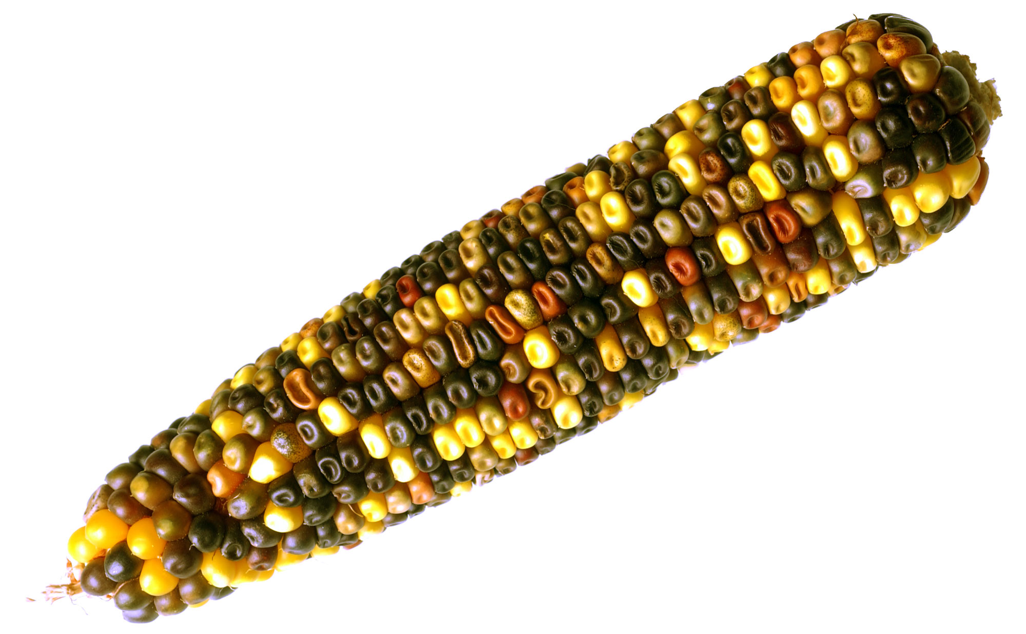
Multicolored ear of corn. Photo by Carl Davies/CSIRO (Wikimedia Commons, Creative Commons Attribution 3.0 Unported license, image cropped and resized).
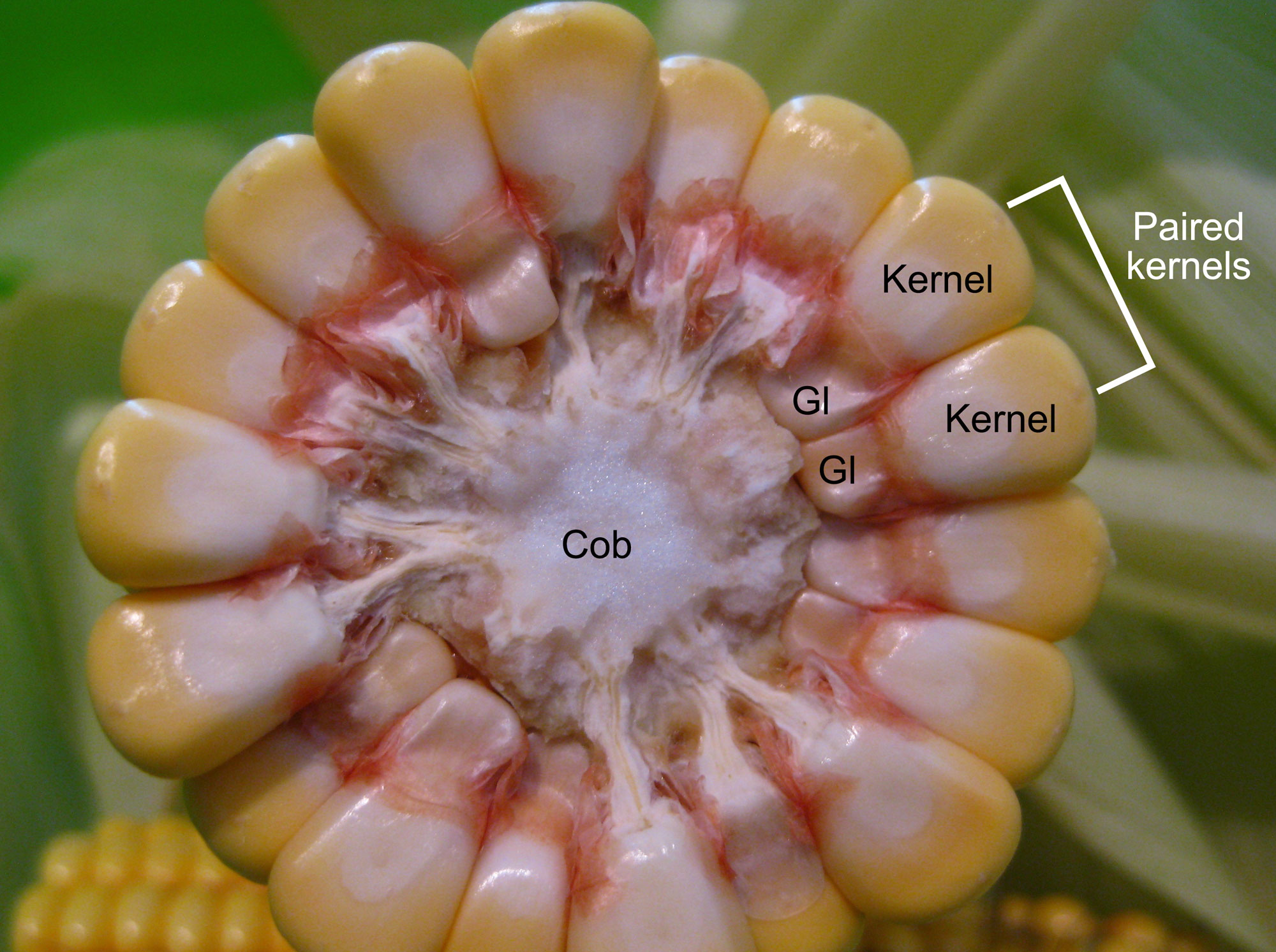
An ear of corn in cross section (cut horizontally) to show the structure. This cob has 18 rows (9 pairs) of kernels. Gl = glume. Photo by Picdrome Public Domain Pictures on flickr (CC0 1.0 Universal/public domain dedication).
A kernel consists of a maize embryo and endosperm, which serves as food for the embryo once it begins growing into a seedling. The endosperm is divided into two types: the harder horny endosperm and the softer floury endosperm. The outer layer of the endosperm is the aleurone layer. The embryo and endosperm are surrounded by a thin pericarp, or fruit wall. The glumes do not cover the fruit when it is mature.
The maize embryo is complex. The upper end of the embryo is the plumule (young stem with young leaves), which is covered by a coleoptile, or protective sheath. The radicle (embryonic root) at the opposite end of the embryo, is also protected by a sheath, the coleorhiza. A cotyledon (seed leaf) called a scutellum is attached to the embryo between the plumule and the radicle. The scutellum shuttles nutrition from the endosperm to the embryo to provide energy during seed germination.
The region of the embryo between the radicle and the attachment point of the scutellum is the hypocotyl, and the region between the plumule and the attachment point of the scutellum is the mesocotyl. Seminal roots, some of the first roots of the young maize plant, may develop on the hypocotyl. The mesocotyl elongates during germination, helping to push the shoot of the developing seedling through the soil.
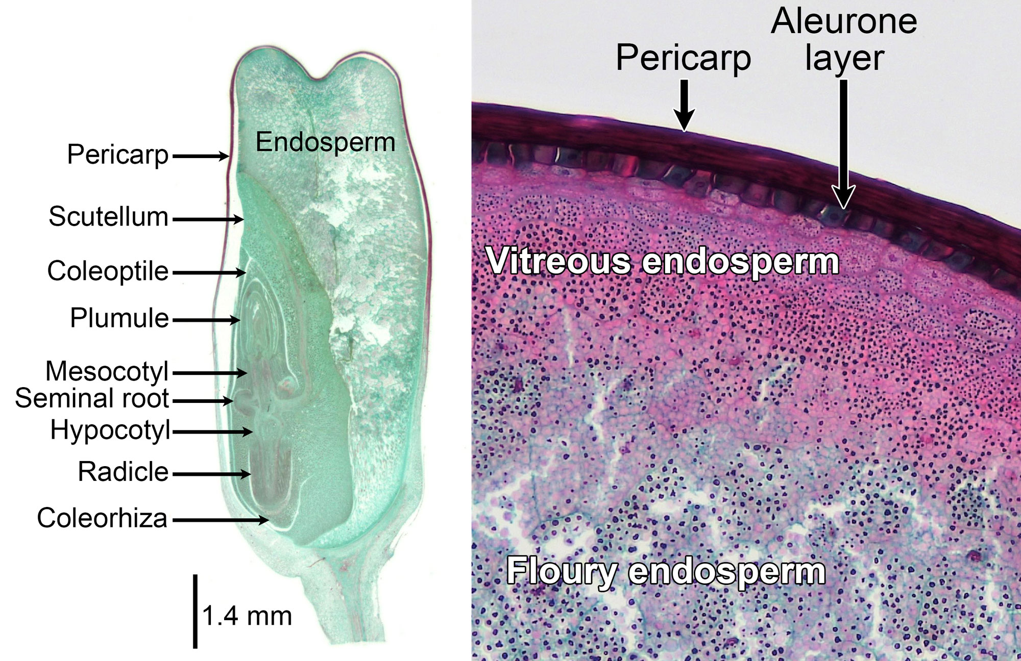
Kernel (grain or caryopsis) of maize (Zea mays). Left: Longitudinal section of a mature kernel. Right: Detail showing the pericarp (fruit wall), aleurone layer, and endosperm. Maize has both hard endosperm (called vitreous endosperm) and soft endosperm (called floury endosperm). Left photo by Jon Houseman and Matthew Ford (Wikimedia Commons, Creative Commons Attribution-ShareAlike 4.0 International license). Right photo by Conor Fitzgerald, 2019 (Berkshire Community College Bioscience Image Library on flickr, public domain). Images cropped, resized, and labeled.
Resources
Additional resources from the Paleontological Research Institution
Digital Encyclopedia of Ancient Life: Introduction to Vascular Plant Structure: https://www.digitalatlasofancientlife.org/learn/embryophytes/tracheophytes/
Earth@Home: Evolution: Grass morphology and anatomy: https://evolution.earthathome.org/grasses/morphology/
Earth@Home: Evolution: Morphology and anatomy of grasses in Tribe Andropogoneae: https://evolution.earthathome.org/grasses/andropogoneae/morphology/
Other websites
Glossary of botanical terms used in Poaceae (Flora of China online): http://flora.huh.harvard.edu/floradata/002/vol22/poaceaeglossary.htm
Poaceae (Graminae) (University of Hawai'i): http://www.botany.hawaii.edu/faculty/carr/po.htm
Scientific articles and books
Clark, L. G., and E. A. Kellogg. In Flora of North America Editorial Committee, Flora of North America North of Mexico vol. 24. Poaceae. Accessed online at: http://floranorthamerica.org/Poaceae
Esau, K. 1977. Anatomy of seed plants, 2nd. ed. John Wiley and Sons, New York.
Evert, R. F., and S. E. Eichhorn. 2013. Raven Biology of Plants, 8th ed. W.H. Freeman and Company Publishers, New York, New York.
Kellogg, E. A. 2015. Flowering Plants, Monocots, Poaceae. The Families and Genera of Vascular Plants 13, 416 pp. (K. Kubitzki, ed.). Springer International Publishing, Switzerland. https://doi.org/10.1007/978-3-319-15332-2_1
Twiss, P. C., E. Suess, and R. M. Smith. 1969. Morphological classification of grass phytoliths. Proceedings of the Soil Science Society of America 33: 109-115.
Weatherwax, P. 1916. Morphology of the flowers of Zea mays. Bulletin of the Torrey Botanical Club 43: 127-144. https://doi.org/10.2307/2479625